
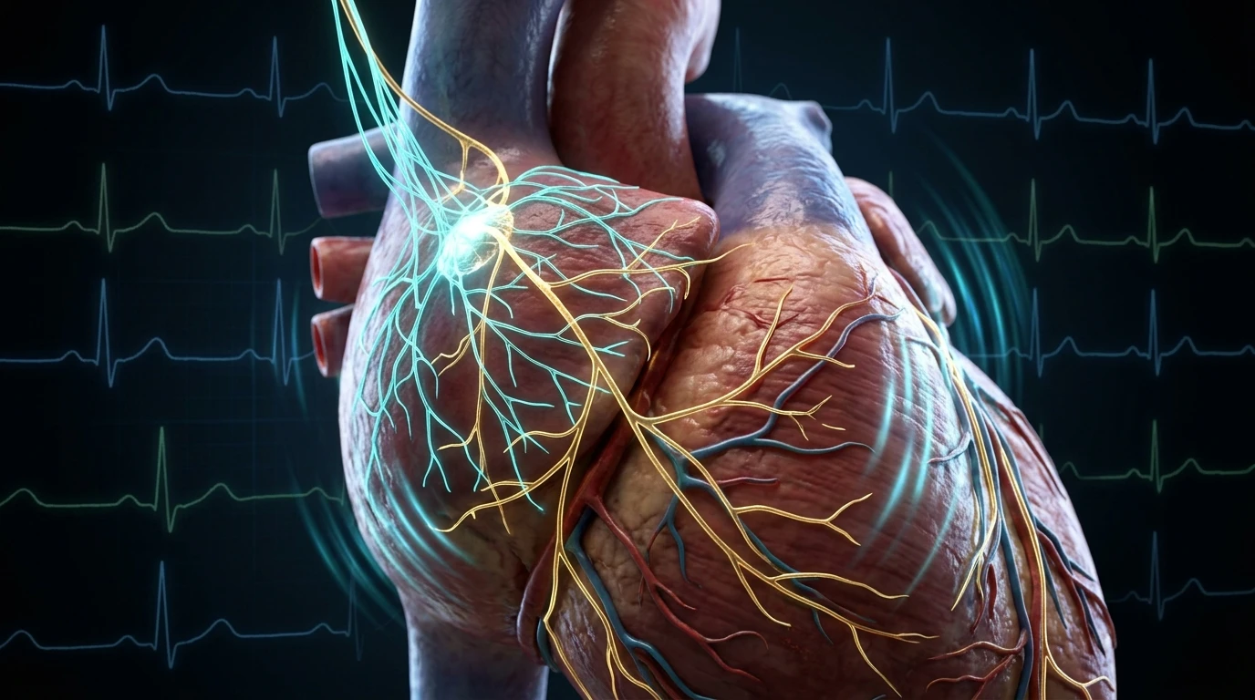
1. The Intrinsic SA Node Rate & The Vagal Brake Concept
Most individuals assume that a resting heart rate of 65 or 70 beats per minute represents the baseline natural rhythm of cardiac muscle tissue. In reality, the human cardiac pacemaker—the sinoatrial (SA) node—possesses an intrinsic spontaneous depolarization rate between 100 and 110 beats per minute.
If the heart were surgically denervated (as occurs during heart transplantation), the resting pulse immediately jumps to approximately 105 BPM. The only reason a healthy resting human heart beats at 60 to 75 BPM is due to continuous, tonic, inhibitory restraint delivered by the parasympathetic nervous system via Cranial Nerve X. In neurocardiology, this physiological phenomenon is termed the Vagal Brake.
When you stand up, take a brisk step, or need to direct attention to an incoming task, the brainstem does not necessarily dump adrenaline into the bloodstream. Instead, it simply eases pressure off the vagal brake, allowing the heart rate to elevate smoothly within milliseconds. Once the transient demand passes, re-applying the vagal brake instantly drops the pulse back down. When this brake fails, cardiovascular instability and sudden panic sensations inevitably follow.
2. Cellular Mechanisms: Acetylcholine, M2 Receptors & Hyperpolarization
The speed and elegance of the vagal brake are governed by precise biophysical and electrophysiological events at the junction between parasympathetic nerve terminals and SA nodal pacemaker cells:
- Foundational Neurobiology: Review our clinical analysis on the physiological sigh and autonomic anxiety reset.
- Acetylcholine Release: Postganglionic vagal motor neurons release acetylcholine (ACh) directly into the synaptic cleft adjacent to cardiac myocytes in the sinoatrial node.
- M2 Muscarinic Receptor Binding: Acetylcholine binds to G-protein coupled M2 muscarinic cholinergic receptors on the cardiac cell membrane.
- Potassium Influx (IKACh Activation): Activation of the M2 receptor dissociates the G-protein beta-gamma subunit, directly opening specialized acetylcholine-activated potassium channels (IKACh). Efflux of positively charged potassium ions hyperpolarizes the membrane, driving the resting membrane potential further away from the firing threshold.
- Inhibition of Funny Currents (If): Concurrently, the alpha subunit of the Gi protein inhibits adenylate cyclase, decreasing intracellular cyclic AMP (cAMP). This directly slows the activation of hyperpolarization-activated cyclic nucleotide-gated (HCN) channels responsible for the spontaneous pacemaker "funny current" (If).
Because these chemical reactions operate locally at the receptor without requiring hormone circulation through the bloodstream, vagal cardio-inhibition takes effect within a single cardiac cycle (under 250 milliseconds). Sympathetic stimulation via circulating norepinephrine, by comparison, requires 3 to 5 seconds to achieve full peak effect.
3. Clinical Manifestations: IST, Post-Viral POTS & Nocturnal Spikes
When the vagal brake becomes impaired through chronic sympathetic allostatic load, autonomic neuropathy, or post-viral inflammatory damage, patients present with distinct clinical syndromes:
Inappropriate Sinus Tachycardia (IST)
Patients with IST experience unexplained resting heart rates consistently exceeding 100 BPM, or disproportionate surges (>130 BPM) with minimal exertion or minor postural shifts, despite structurally normal echocardiograms. Research demonstrates that over 60% of IST cases stem primarily from a loss of intrinsic parasympathetic vagal tone rather than excessive catecholamine production.
Nocturnal Heart Rate Spikes
Under normal conditions, sleep onset and non-REM deep sleep are characterized by profound vagal dominance, plunging heart rate to its lowest daily nadir. Patients with vagal brake failure frequently wake up gasping at 2:00 AM or 3:00 AM with a racing pulse (110-130 BPM), mistakenly believing they have experienced a panic attack or cardiac emergency.
4. Differential Comparison: Normal Vagal Brake vs. Brake Failure
| Clinical Parameter | Intact Vagal Brake (Healthy ANS) | Vagal Brake Dysfunction / Failure |
|---|---|---|
| Resting Heart Rate | 60 – 75 BPM | 95 – 115+ BPM (at complete rest) |
| Respiratory Sinus Arrhythmia | Pronounced (heart accelerates on inhale, slows on exhale) | Blunted or absent (metronomic pulse) |
| Heart Rate Variability (RMSSD) | High (>40 ms, age-adjusted) | Severely depressed (<15 ms) |
| Response to Mild Stress | Smooth 10-15 BPM increase via brake release; rapid recovery | Sharp 30-50 BPM spikes; prolonged recovery time |
| Nocturnal Sleep Nadir | Drop of 15-20% below daytime baseline | Elevated flatline; nocturnal adrenergic surges |
| Underlying Driver | Adaptive autonomic flexibility | Inflammation of NTS, cholinergic deficiency, or post-viral vagopathy |
5. Evidence-Based Protocols to Restore Cardio-Vagal Inhibition
Re-establishing parasympathetic cardio-inhibition requires daily neuroplastic retraining through physiological reflexes that bypass cortical anxiety loops:
1. Baroreflex Retraining via Resonant Breathing
Breathing at approximately 5.5 to 6 breaths per minute (typically 4.5 seconds in, 5.5 seconds out) aligns respiratory oscillations with the intrinsic 0.1 Hz Mayer waves of the vascular baroreflex. This resonance dramatically stimulates baroreceptors in the carotid sinus and aortic arch, forcing the brainstem to re-engage acetylcholine output to the SA node.
2. Trigeminal-Vagal Dive Reflex Activation
Applying ice-cold water (under 15°C / 59°F) directly to the ophthalmic branch (V1) of the trigeminal nerve on the upper cheeks and forehead while holding a calm breath activates the mammalian dive reflex. The sensory input relays directly to the NTS, triggering an immediate, involuntary parasympathetic cardio-deceleration within 10 to 20 seconds.
3. Cholinergic Nutrient Optimization
Synthesis of acetylcholine requires adequate dietary intake of choline (such as citicoline or alpha-GPC) alongside vitamin B1 (thiamine) and acetyl-CoA derived from healthy mitochondrial carbohydrate metabolism.
Frequently Asked Questions (Clinical FAQ)
What is the natural intrinsic heart rate without the vagus nerve?
The intrinsic pacemaker rate of the human sinoatrial (SA) node is between 100 and 110 beats per minute. Any resting heart rate lower than this is actively held down by parasympathetic vagal acetylcholine release.
How does vagal brake dysfunction trigger panic attacks?
When the vagal brake fails, resting heart rate spikes suddenly without a conscious psychological trigger. The brain's interoceptive cortex interprets this rapid cardiac acceleration as an impending life threat, creating a secondary wave of panic, hyperventilation, and catastrophic thinking.
Can dehydration cause vagal brake failure?
Dehydration reduces venous return and stroke volume. To maintain blood pressure and cardiac output, the brainstem must pull off the vagal brake and activate sympathetic drive, causing resting and orthostatic tachycardia.
What laboratory tests evaluate the cardiac vagal brake?
Clinical autonomic testing labs utilize 24-hour Holter monitoring, high-frequency (HF) spectral analysis of HRV, the Valsalva maneuver, and deep breathing heart rate variation tests (E:I ratio).
Are beta-blockers the only treatment for a failed vagal brake?
While beta-blockers (like propranolol or metoprolol) and funny-channel inhibitors (ivabradine) are standard pharmacological tools to control heart rate, they act primarily as external dampeners. Restoring endogenous vagal tone through baroreflex retraining, cold face immersion, and sleep optimization addresses the root autonomic pathology.
Does vagal brake failure damage the heart muscle?
Inappropriate sinus tachycardia originating from a failed vagal brake rarely damages the heart muscle directly, as cardiac output and ventricular function typically remain preserved. However, the continuous high pulse causes immense physical exhaustion, exercise intolerance, and severe cognitive strain.
Scientific References & Clinical Citations
- The Vagal Brake: A Psychophysiological Marker of Emotion Regulation — Psychophysiology Journal (2007). [PubMed / Study Link]
- Inappropriate Sinus Tachycardia: Pathophysiology and Contemporary Management — Journal of the American College of Cardiology (2020). [PubMed / Study Link]
- Cardiovascular Autonomic Regulation: From Basic Mechanisms to Clinical Practice — Circulation Research (2021). [PubMed / Study Link]
- Muscarinic M2 Receptor Signaling and Cardiac Pacemaker Control — Physiological Reviews (2018). [PubMed / Study Link]