
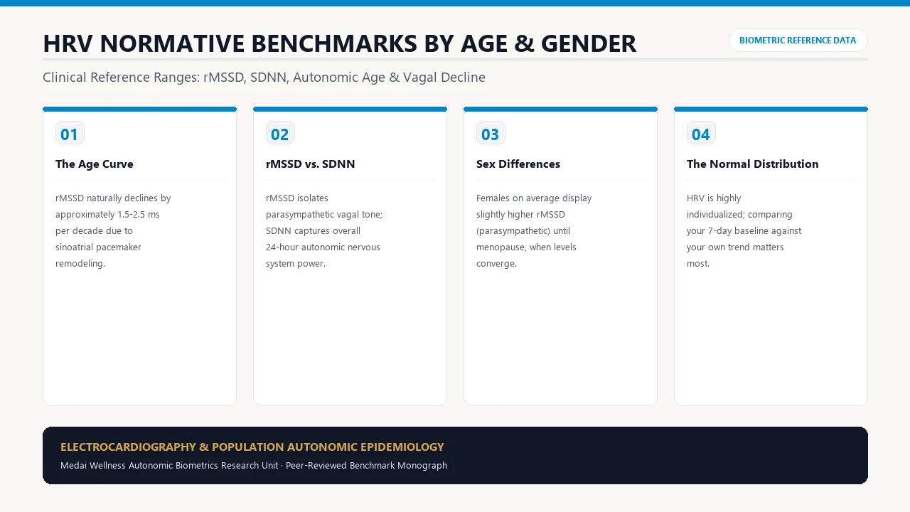
1. What HRV Truly Measures: Sympathovagal Balance
A common misconception is that a healthy resting heart beats with metronomic regularity. In reality, a rigid, unchanging heartbeat is a marker of autonomic exhaustion, cardiovascular stiffness, and increased all-cause mortality risk. In a healthy human, the time elapsed between consecutive cardiac contractions (R-R intervals) fluctuates constantly by tens to hundreds of milliseconds.
This continuous beat-to-beat variability is driven by the dynamic tension between the sympathetic nervous system (accelerating heart rate via norepinephrine at beta-1 receptors) and the parasympathetic nervous system (decelerating heart rate via vagal acetylcholine at muscarinic M2 receptors). Heart Rate Variability (HRV) serves as the most accurate non-invasive window into central autonomic nervous system adaptability.
2. Normative HRV Benchmark Tables (RMSSD & SDNN by Age and Sex)
The following normative data synthesizes findings from large-scale multi-cohort studies, including the Task Force of the European Society of Cardiology, the American College of Cardiology, and comprehensive 24-hour ambulatory monitoring datasets (incorporating over 25,000 subjects):
| Age Bracket | Male (25th – 75th Percentile) | Male Median | Female (25th – 75th Percentile) | Female Median | Elite Endurance Athlete Range |
|---|---|---|---|---|---|
| 20 – 29 Years | 42 – 76 ms | 56 ms | 39 – 72 ms | 53 ms | 95 – 140+ ms |
| 30 – 39 Years | 32 – 58 ms | 44 ms | 31 – 56 ms | 42 ms | 75 – 115 ms |
| 40 – 49 Years | 24 – 45 ms | 34 ms | 24 – 43 ms | 33 ms | 58 – 90 ms |
| 50 – 59 Years | 18 – 34 ms | 25 ms | 18 – 33 ms | 25 ms | 42 – 70 ms |
| 60 – 69 Years | 14 – 27 ms | 19 ms | 14 – 26 ms | 19 ms | 32 – 52 ms |
| 70+ Years | 10 – 21 ms | 15 ms | 11 – 22 ms | 16 ms | 24 – 40 ms |
3. RMSSD vs. SDNN: Selecting the Right Physiological Metric
| Metric | Mathematical Derivation | Primary Autonomic Driver | Clinical Use Case |
|---|---|---|---|
| RMSSD (ms) | Root Mean Square of Successive Differences | Parasympathetic / Vagal Tone exclusively | Short-term recovery, sleep quality, daily readiness |
| SDNN (ms) | Standard Deviation of all normal-to-normal (NN) intervals | Combined Sympathetic + Parasympathetic + Circadian | 24-hour Holter monitoring; overall cardiovascular prognosis |
| pNN50 (%) | Percentage of successive NN intervals differing by >50ms | Parasympathetic / Vagal cholinergic output | Complementary metric to RMSSD in resting state testing |
4. The Neurobiology of Age-Related HRV Decline
Across population datasets, HRV displays an unyielding downward slope with advancing chronological age. Three physiological mechanisms explain this progression:
- Structural Arterial Remodeling: As central arteries lose elastin and accrue collagen cross-linking, the transmission of pulse pressure waves to the carotid sinus and aortic baroreceptors becomes dampened. Blunted baroreflex mechanosensation directly curtails efferent vagal burst activity.
- Sinoatrial Node Atrophy: Pacemaker cells in the sinoatrial node naturally diminish in quantity by ~10% per decade, reducing the responsiveness of pacemaker tissue to acetylcholine.
- Central Cholinergic Decline: Nucleus Ambiguus and dorsal motor nucleus output declines with systemic neuroinflammation and cerebral microvascular aging.
5. Measurement Protocols: Clinical ECG vs. Chest Straps vs. Wearables
To avoid misinterpreting erratic readings, understanding hardware limitations is critical:
- Gold Standard (ECG & Chest Straps): A 12-lead clinical ECG or a dedicated bipolar chest strap (such as the Polar H10) records actual myocardial depolarization voltages (R-waves) at 1,000 Hz, with near-zero error.
- Optical PPG Wearables (Oura, Whoop, Apple Watch): Use photoplethysmography to detect capillary pulse waves. While daytime PPG is susceptible to motion artifacts, nocturnal PPG measurements taken during slow-wave sleep achieve 95% to 98% concordance with ECG for RMSSD tracking.
6. Evidence-Based Protocols to Elevate Baseline Vagal Tone
While chronological age sets the reference boundary, lifestyle inputs can shift an individual from the 25th percentile into the 90th percentile for their age bracket:
- Zone 2 Aerobic Conditioning: 150 to 180 minutes weekly of continuous cardiovascular training at 60-70% max HR stimulates cardiac eccentric hypertrophy, expands stroke volume, and elevates resting vagal braking.
- Nocturnal Alcohol Elimination: Even moderate alcohol consumption (1-2 drinks) suppresses nighttime RMSSD by 30% to 60% by elevating sympathetic tone and disrupting slow-wave sleep.
- Resonance Frequency Breathing: Practicing paced diaphragmatic breathing at 5.5 to 6.0 breaths per minute for 15 minutes daily trains the baroreflex and produces sustained elevations in baseline RMSSD.
7. Frequently Asked Clinical Questions (FAQ)
Why did my HRV suddenly drop by 40% overnight?
Acute drops in HRV almost always indicate acute autonomic stressors: the onset of a viral infection (often appearing 24-48 hours before subjective symptoms), alcohol consumption, late heavy dining, intense unrecovered muscular trauma, or severe emotional distress.
Is a higher HRV always better?
Within normal physiological ranges, higher HRV reflects superior parasympathetic recovery and autonomic elasticity. However, sudden spikes above 150 ms in non-athletes frequently represent arrhythmias like premature ventricular contractions (PVCs) that trick optical algorithms into calculating false variability.
How does hydration affect HRV measurements?
Dehydration reduces circulating blood volume, which forces the heart to beat faster to maintain cardiac output. This compensatory sympathetic activation lowers resting HRV. Maintaining adequate fluid and electrolyte balance is essential for accurate baseline measurements.
Should I compare my HRV number to my friends or family?
No. HRV is intensely individual, influenced by genetics, left ventricular chamber volume, and baseline vagal nerve anatomy. The only clinically valid comparison is comparing your daily reading against your own 14-day rolling baseline.
Can cold water immersion improve HRV?
Yes. Brief cold water exposure (2-3 minutes at 50-55°F) triggers the mammalian dive reflex, causing acute peripheral vasoconstriction followed by a sustained rebound in parasympathetic vagal output that elevates post-exposure RMSSD.
8. Peer-Reviewed Citations
- Shaffer F, Ginsberg JP. An Overview of Heart Rate Variability Metrics and Norms. Frontiers in Public Health. 2017;5:258. PMID: 29034226.
- Task Force of the European Society of Cardiology and the North American Society of Pacing and Electrophysiology. Heart rate variability: standards of measurement, physiological interpretation and clinical use. Circulation. 1996;93(5):1043-1065. PMID: 8598068.
- Umetani K, Singer DH, McCraty R, Atkinson M. Twenty-four hour time domain heart rate variability: Normalized values for healthy adults by age and sex. Journal of the American College of Cardiology. 1998;31(3):593-601. PMID: 9502641.
- Nunan D, Sandercock GR, Brodie DA. A quantitative systematic review of normal values for short-term heart rate variability in healthy adults. Pacing and Clinical Electrophysiology. 2010;33(11):1407-1417. PMID: 20663071.