
1. The Neuroendocrinology of the Diurnal Cortisol Curve
Cortisol, the primary glucocorticoid produced by the zona fasciculata of the human adrenal cortex, is vital for maintaining vascular tone, carbohydrate and lipid metabolism, immune modulation, and circadian entrainment. Cortisol does not circulate at a fixed baseline; its secretion is strictly pulsatile and circadian.
This rhythm is driven by the master circadian clock—the suprachiasmatic nucleus (SCN) within the anterior hypothalamus. The SCN transmits neural and humoral signals to the paraventricular nucleus (PVN), dictating the release of corticotropin-releasing hormone (CRH) and arginine vasopressin (AVP), which in turn stimulate anterior pituitary adrenocorticotropic hormone (ACTH) pulsatility.
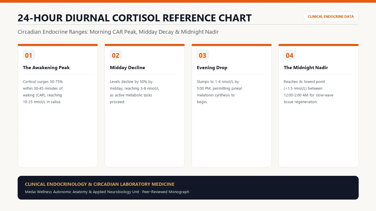
2. 24-Hour Reference Ranges Chart (Salivary Free Cortisol & Serum)
Below is the definitive clinical reference chart synthesizing reference intervals established by major endocrine testing institutions (LabCorp, Quest Diagnostics, and the Endocrine Society guidelines):
| Timepoint / Circadian Phase | Salivary Free Cortisol (nmol/L) | Salivary Free Cortisol (ng/mL) | Serum Total Cortisol (μg/dL) | Physiological Objective |
|---|---|---|---|---|
| Waking Baseline (0 min) | 10.2 – 27.3 nmol/L | 3.7 – 9.9 ng/mL | 10.0 – 20.0 μg/dL | Initial arousal; termination of melatonin dominance |
| Peak CAR (+30-45 min) | 15.0 – 38.5 nmol/L | 5.4 – 14.0 ng/mL | 15.0 – 25.0 μg/dL | Maximal gluconeogenesis; cognitive anticipation |
| Midday (12:00 PM) | 3.5 – 10.8 nmol/L | 1.3 – 3.9 ng/mL | 6.0 – 14.0 μg/dL | Metabolic energy maintenance without anxiety |
| Afternoon (4:00 - 5:00 PM) | 2.0 – 6.5 nmol/L | 0.7 – 2.4 ng/mL | 3.0 – 10.0 μg/dL | Steady circadian decline; energy stabilization |
| Evening Nadir (10:00 - 11:00 PM) | < 2.0 nmol/L | < 0.7 ng/mL | < 3.5 μg/dL | Permissive threshold for pineal melatonin synthesis & slow-wave sleep |
3. The Cortisol Awakening Response (CAR): Clinical Significance
The Cortisol Awakening Response (CAR) represents an acute 50% to 75% surge in free cortisol within 30 to 45 minutes following eyes-open consciousness. Unlike the passive basal nocturnal rise (which begins around 3:00 AM), the CAR is an active, anticipatory neuro-endocrine reflex mediated by a direct polysynaptic pathway connecting the hippocampus, prefrontal cortex, and SCN to the adrenal glands.
An optimal CAR serves two vital functions: it rapidly upregulates hepatic gluconeogenesis to deliver glucose to the waking brain, and it sets the phase of peripheral circadian clocks located in liver, skeletal muscle, and adipose tissue.
4. Pathological Diurnal Curves: Burnout, Inversion & Hypervigilance
| Curve Phenotype | Diagnostic Hallmarks | Clinical Etiology | Common Symptoms |
|---|---|---|---|
| Flattened / Exhausted Curve | Blunted CAR (<10 nmol/L); flat slope across the day | Chronic HPA axis burnout, PTSD, long-term systemic inflammation | Unrefreshing sleep, severe morning fatigue, chronic pain, brain fog |
| Elevated Night Curve | Normal morning CAR; evening cortisol >4.0 nmol/L | Sympathetic nervous system hyperarousal, nighttime digital exposure | Sleep-onset insomnia, 3 AM awakenings, racing heart, visceral fat gain |
| Inverted Circadian Curve | Low morning waking; paradoxical spike at 10 PM | Shift work, severe circadian desynchrony, adrenal receptor dysregulation | Daytime lethargy, nighttime hyper-focus ("wired and tired" state) |
5. Diagnostic Modalities: Saliva vs. Serum vs. 24h Urine vs. DUTCH
Accurate clinical evaluation requires choosing the appropriate biofluid. In routine clinical practice, 4-point or 5-point salivary free cortisol collection represents the gold standard for mapping the circadian curve. Salivary cortisol reflects the free, unbound fraction that exerts biological effects on glucocorticoid receptors, avoiding the venipuncture anxiety that artificially spikes serum cortisol levels.
6. Clinical Strategies to Restore a Blunted Cortisol Slope
Restoring a healthy diurnal slope requires targeted behavioral and circadian interventions:
- Morning Outdoor Lux Exposure: View 10,000+ lux of natural morning sunlight within 30 minutes of waking for 15 to 20 minutes to reset the SCN and amplify the CAR.
- Caffeine Timing Protocol: Delay first caffeine intake by 90 to 120 minutes post-waking to allow endogenous adenosine clearance and prevent morning CAR blunting.
- Evening Light Attenuation: Eliminate high-kelvin overhead blue light 2 hours before bed; keep evening salivary cortisol below the 2.0 nmol/L threshold.
- Phosphatidylserine Modulation: For patients with elevated evening cortisol, 300 to 400 mg of standardized phosphatidylserine at 6:00 PM dampens nocturnal ACTH hyper-secretion without depressing morning tone.
7. Frequently Asked Clinical Questions (FAQ)
Why is high evening cortisol associated with visceral belly fat?
Visceral intra-abdominal adipose tissue contains up to 4 to 5 times higher density of glucocorticoid receptors (GR) and 11β-HSD1 enzymes than subcutaneous fat. When cortisol remains elevated at night in the presence of insulin, it selectively activates lipoprotein lipase in mesenteric fat depots, mobilizing triglycerides into deep visceral stores.
Can intense exercise in the evening ruin my cortisol curve?
Yes. High-intensity interval training (HIIT) or heavy weightlifting performed within 3 hours of sleep can trigger a 30% to 50% spike in salivary free cortisol, delaying the nocturnal nadir and impairing slow-wave sleep. Reserve intense workouts for morning or early afternoon, keeping evening exercise limited to light walking or gentle mobility work.
How do oral contraceptives affect cortisol lab results?
Exogenous estrogens increase hepatic synthesis of Cortisol-Binding Globulin (CBG). As a result, total serum cortisol blood tests appear artificially elevated, while salivary free cortisol remains accurate and reflective of genuine unbound tissue exposure.
What is the difference between Addison's disease and HPA axis fatigue?
Addison's disease is an autoimmune or structural destruction of the adrenal cortex resulting in near-zero cortisol production (a medical emergency). Functional HPA axis dysregulation (burnout) involves hypothalamic and pituitary receptor desensitization and altered central tone, where the adrenal glands remain structurally intact.
How long does it take to re-establish a normal cortisol curve?
Clinical observation demonstrates that strict adherence to circadian light hygiene, sleep scheduling, stress downregulation, and nutritional timing produces noticeable curve improvement within 6 to 12 weeks on follow-up 4-point salivary testing.
8. Peer-Reviewed Citations & Clinical Guidelines
- Clow A, Hucklebridge F, Stalder T, Evans P, Thorn L. The cortisol awakening response: More than a measure of HPA axis function. Neuroscience & Biobehavioral Reviews. 2010;35(1):97-103. PMID: 20045431.
- Nieman LK, Biller BM, Findling JW, Newell-Price J, Savage MO, Stewart PM, Montori VM. The diagnosis of Cushing's syndrome: An Endocrine Society Clinical Practice Guideline. The Journal of Clinical Endocrinology & Metabolism. 2008;93(5):1526-1540. PMID: 18334580.
- Hellhammer DH, Wüst S, Kudielka BM. Salivary cortisol as a biomarker in social science research. Psychoneuroendocrinology. 2009;34(2):163-171. PMID: 19095172.
- Adam EK, et al. Diurnal cortisol slopes and mental and physical health outcomes: A systematic review and meta-analysis. Psychoneuroendocrinology. 2017;83:25-41. PMID: 28578301.