
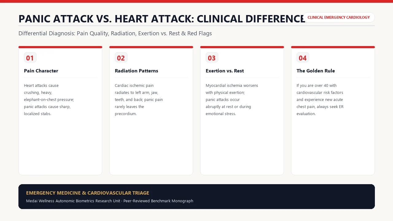
1. Pathophysiological Foundations: Ischemia vs. Sympathetic Storm
Distinguishing between an acute panic attack and an acute myocardial infarction (MI) is one of the most frequent and high-stakes diagnostic challenges in emergency medicine. Annually, over 6 million patients present to United States emergency departments with acute chest pain; between 15% and 25% of these individuals are experiencing acute panic disorder rather than coronary syndrome.
Despite their shared sensations of impending doom and tachycardia, their biological drivers are entirely distinct:
- Myocardial Infarction (Heart Attack): Occurs when an atherosclerotic plaque in a coronary artery ruptures, inducing localized platelet aggregation and thrombosis. This occludes myocardial blood supply, causing cellular hypoxia, lactic acidosis, and myocardial tissue necrosis.
- Panic Attack: Driven by an abrupt, unprovoked activation of the central fear circuit centered in the amygdala. The locus coeruleus floods the system with norepinephrine while the adrenal medulla discharges adrenaline. This triggers extreme peripheral vasoconstriction, intercostal muscle spasm, and hyperventilation-induced respiratory alkalosis.
2. Comprehensive Differential Diagnostic Matrix (12 Clinical Markers)
| Clinical Feature | Panic Attack (Autonomic Surge) | Heart Attack (Myocardial Infarction) |
|---|---|---|
| Onset Characteristic | Abrupt, sudden onset, often at rest without physical trigger | Gradual progression or sudden onset; frequently triggered by physical exertion or stress |
| Peak Severity | Reaches maximum intensity within 10 minutes | Steadily builds or remains unrelenting over hours |
| Total Duration | Resolves gradually within 20 to 30 minutes | Lasts longer than 20 minutes; does not resolve spontaneously |
| Nature of Chest Discomfort | Sharp, stabbing, burning, or tender intercostal aches | Heavy, crushing, squeezing pressure; "elephant on the chest" |
| Location of Pain | Localized to a small, pinpointable spot on chest wall | Diffuse retrosternal pain across the center/left chest |
| Pain Radiation | Rarely radiates; stays confined to chest or neck | Frequently radiates to left arm, shoulder, jaw, neck, or back |
| Effect of Deep Breathing | Often worsens pain due to intercostal muscle expansion | Unchanged by respiration or body position |
| Palpation (Pressing on Chest) | Often tender to touch (costochondritis or muscle spasm) | Deep visceral pain; not reproducible by pressing chest wall |
| Neurological Sensations | Perioral tingling, finger numbness (carpopedal spasm) | Profound presyncope, genuine loss of consciousness, cold clamminess |
| Response to Rest | Distraction or physiological breathing reduces symptoms | Rest does not alleviate symptoms if artery is fully occluded |
| Sweating Characteristic | Sweaty palms or generalized autonomic perspiration | Profuse, cold, drenching diaphoresis with pale/ashen skin |
| Age & Risk Factor Profile | Common across all ages; highest prevalence in 18-35 | Significantly higher in adults >45, smokers, diabetics, hypertensives |
3. Pain Characteristics & Anatomical Radiation Patterns
The neuroanatomical sensory wiring of the chest provides critical diagnostic clues. The visceral sensory nerves of the myocardium travel along thoracic sympathetic afferents (T1 to T4 spinal segments). Because these dermatomes share dorsal horn pathways with somatic sensations from the left arm, shoulder, and jaw, the cerebral cortex misinterprets myocardial ischemia as radiating pain in the left upper extremity and mandible.
In contrast, panic attacks stimulate somatic intercostal nerve endings through violent chest wall hyper-expansion and rapid breathing, resulting in superficial, reproducible musculoskeletal sharp pains.
4. Temporal Dynamics: Peak Intensity and Resolution Durations
A crucial differentiator is the symptom trajectory over time. In a panic attack, circulating epinephrine and norepinephrine follow rapid hepatic and renal clearance kinetics. As a result, symptoms hit an explosive zenith within 8 to 10 minutes and steadily fade as the parasympathetic branch re-engages.
In acute coronary syndrome, ischemic tissue damage is progressive. Unless coronary perfusion is restored mechanically via percutaneous coronary intervention (PCI / stent) or fibrinolysis, cardiac pain remains unrelenting or intensifies over hours.
5. Sex-Specific Variations: Atypical Presentation in Women
Warning: Atypical Heart Attacks in Women
Medical literature demonstrates that over 40% of women presenting with acute myocardial infarction do not experience classic crushing retrosternal chest pressure. Instead, female heart attack symptoms frequently manifest as:
- Sudden, overwhelming, unexplainable physical exhaustion or weakness
- Shortness of breath (dyspnea) without prominent chest pain
- Nausea, vomiting, indigestion, or epigastric discomfort mimicking acid reflux
- Dull, aching pain radiating between the shoulder blades or into the throat and lower jaw
- Lightheadedness or near-syncope
Because these symptoms closely resemble somatic anxiety or gastrointestinal distress, women are statistically more likely to experience delayed emergency department arrival.
6. Emergency Department Diagnostic Workup: ECG & Troponins
When presenting to an emergency department with acute chest pain, physicians perform an immediate two-part clinical objective evaluation:
- 12-Lead Electrocardiogram (ECG): Recorded within 10 minutes of arrival. Detects ST-segment elevation (STEMI), T-wave inversions, ST depressions (NSTEMI), or ischemic bundle branch blocks. In a panic attack, the ECG typically shows sinus tachycardia with normal waveforms.
- High-Sensitivity Cardiac Troponin (hs-cTnI / hs-cTnT): Troponins are structural proteins unique to cardiac myocytes. When myocardial cell membranes rupture due to ischemic necrosis, troponin leaks into the bloodstream. Blood levels are tested on arrival and repeated at 1 and 3 hours. In a panic attack, cardiac troponins remain strictly zero.
7. Frequently Asked Clinical Questions (FAQ)
Can a panic attack trigger a real heart attack?
In individuals with clean coronary arteries, a panic attack does not cause an infarction. However, in individuals with severe pre-existing coronary artery stenosis, the severe catecholamine surge, heart rate acceleration, and acute hypertension can trigger a mismatch between myocardial oxygen demand and supply, precipitating ischemia.
Why do my hands and lips tingle during a panic attack?
Rapid hyperventilation expels carbon dioxide faster than cellular metabolism produces it, leading to respiratory alkalosis (elevated blood pH). This causes serum calcium ions to bind to albumin, inducing mild transient hypocalcemia. This hypocalcemia excites peripheral nerve membranes, producing tingling in the lips, fingertips, and carpopedal muscle stiffness.
What is Takotsubo cardiomyopathy (Broken Heart Syndrome)?
Takotsubo cardiomyopathy is a temporary condition where acute emotional stress floods the left ventricle with massive levels of adrenaline, causing the apex of the heart to balloon outward and stunning heart muscle. It mimics a heart attack on ECG and requires immediate emergency care, but coronary arteries show no obstructive clots.
Should I take an aspirin if I am not sure which one I am having?
If you suspect a heart attack and have no allergy to aspirin or active gastrointestinal bleeding, emergency guidelines recommend chewing one adult 325 mg aspirin (or 2 to 4 low-dose 81 mg baby aspirins) while awaiting emergency medical responders. Chewing allows rapid buccal and gastric absorption to inhibit platelet aggregation.
How can I calm myself down if I know it is just a panic attack?
Perform the Physiological Sigh: two quick nasal inhalations followed by an extended, passive mouth exhalation. Repeat 3 times. Splash cold water on your face to activate the trigeminal-cardiac vagal reflex, and verbally remind yourself: "This is a temporary surge of adrenaline; my heart is structurally sound and this will peak and dissipate within 10 minutes."
8. Peer-Reviewed Citations & Clinical Guidelines
- Gulati M, Levy PD, Mukherjee D, et al. 2021 AHA/ACC/ASE/CHEST/SAEM/SCCT/SCMR Guideline for the Evaluation and Diagnosis of Chest Pain: A Report of the American College of Cardiology/American Heart Association Joint Committee on Clinical Practice Guidelines. Circulation. 2021;144(22):e368-e454. PMID: 34709923.
- Fleet RP, Dupuis G, Marchand A, et al. Panic disorder in emergency department chest pain patients: Prevalence, comorbidity, and cardiovascular risk. Journal of Psychosomatic Research. 1996;41(4):307-320. PMID: 8971661.
- Canto JG, Goldberg RJ, Hand MM, et al. Symptom presentation of women with acute coronary syndromes: Myth vs reality. Archives of Internal Medicine. 2007;167(22):2405-2413. PMID: 18071161.
- Thygesen K, Alpert JS, Jaffe AS, et al. Fourth Universal Definition of Myocardial Infarction (2018). European Heart Journal. 2019;40(3):237-269. PMID: 30165616.