
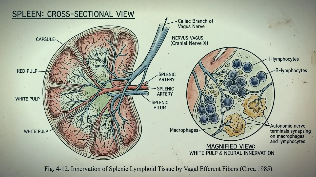
1. The Molecular Discovery: Beyond the Humoral Immune Paradigm
For over a century, immunology viewed the immune system as an autonomous, self-regulating biological network operating entirely via biochemical feedback loops, antibodies, and circulating cytokines. In 2000, a landmark discovery published in Nature by neurosurgeon Dr. Kevin J. Tracey and his team at the Feinstein Institutes for Medical Research overturned this dogma.
While developing a synthetic stroke compound designed to block brain inflammation, researchers discovered that administering tiny, picogram amounts of the drug intracerebroventricularly into the brainstem completely prevented lethal systemic tumor necrosis factor-alpha (TNF-α) production in the liver, spleen, and bloodstream during endotoxemia. Cutting the cervical vagus nerve completely eliminated this protective effect. The Cholinergic Anti-Inflammatory Pathway (CAIP) was born.
The realization that the central nervous system controls systemic immunity via electrical pulses down the vagus nerve established the foundation for modern bioelectronic medicine and autonomic neuro-immunology, closely tied to our clinical research on exercises to raise vagal tone.
2. The Neuroanatomical Circuit: Vagus → Celiac Ganglion → Splenic Nerve
Mapping the anatomical trajectory of the inflammatory reflex revealed an unexpected neuro-vascular architecture. While sensory vagal afferents in the nodose ganglion constantly sense peripheral cytokine concentrations and transmit inflammatory alarms to the Nucleus Tractus Solitarius (NTS), the motor efferent arm follows a precise sequence:
- Brainstem Origin: Efferent motor signals originate in the Dorsal Motor Nucleus of the Vagus (DMNX) and descend through the cervical and subdiaphragmatic vagal trunks.
- The Celiac Ganglion Synapse: The subdiaphragmatic vagus does not directly penetrate the spleen. Instead, cholinergic pre-ganglionic fibers terminate in the celiac-superior mesenteric ganglion complex.
- The Splenic Nerve Transition: In the celiac ganglion, vagal inputs synapse onto post-ganglionic sympathetic neurons, transferring the signal into the splenic nerve. The splenic nerve travels alongside the splenic artery into the red pulp of the spleen, releasing norepinephrine into the lymphoid microenvironment.
3. Choline Acetyltransferase (ChAT+) T-Cells: The Cellular Transducers
A critical mystery remained: if the splenic nerve releases norepinephrine, how does the pathway become "cholinergic" (acetylcholine-mediated)?
In a series of landmark investigations published in Science, Rosas-Ballina et al. identified the missing cellular link: a unique subpopulation of specialized memory T lymphocytes expressing the enzyme choline acetyltransferase (ChAT). These ChAT+ CD4+ T-cells reside in the marginal zone and red pulp of the spleen.
When the splenic nerve fires, norepinephrine binds to β2-adrenergic receptors on these ChAT+ T-cells. This adrenergic signal activates intracellular adenylate cyclase, prompting these T-cells to synthesize, store, and release massive quantities of acetylcholine directly into the splenic parenchyma.
4. The α7 Nicotinic Receptor: Halting NF-κB Nuclear Translocation
Once released by splenic T-cells, acetylcholine diffuses onto adjacent splenic red pulp macrophages, dendritic cells, and monocytes. These immune cells express high densities of the alpha-7 nicotinic acetylcholine receptor (α7 nAChR)—a homopentameric ligand-gated ion channel.
Binding of acetylcholine to the α7 nAChR initiates an immediate intracellular signaling cascade:
- JAK2-STAT3 Activation: Receptor activation triggers rapid phosphorylation of Janus kinase 2 (JAK2), which subsequently phosphorylates Signal Transducer and Activator of Transcription 3 (STAT3).
- Suppression of NF-κB: Phosphorylated STAT3 homodimers translocate to the cell nucleus, physically interfering with and blocking the nuclear translocation of Nuclear Factor kappa B (NF-κB p65).
- Cytokine Gene Silencing: Because NF-κB is the master transcriptional switch for inflammatory cytokines, its blockade halts the transcription of genes encoding TNF-α, Interleukin-1 beta (IL-1β), Interleukin-6 (IL-6), and High Mobility Group Box 1 (HMGB1).
5. Clinical Implications: Autoimmunity, POTS, Long COVID & Sepsis
When the cholinergic anti-inflammatory pathway is chronically impaired, unchecked low-grade neuroinflammation ravages peripheral tissues. In conditions such as POTS, Long COVID, Rheumatoid Arthritis, and Inflammatory Bowel Disease (IBD), patients exhibit blunted vagal nerve activity and elevated systemic circulating TNF-α and IL-6 levels.
Clinical trials using surgically implanted vagus nerve stimulators in patients with drug-resistant rheumatoid arthritis and Crohn's disease demonstrated up to a 70% reduction in circulating TNF-α levels and substantial clinical remission, proving the viability of bioelectronic immunosuppression.
6. Methods to Activate the Pathway: VNS, Breathing & Choline Agonists
Modern non-pharmacological therapies can potently stimulate this anti-inflammatory pathway:
- Transcutaneous Auricular Vagus Nerve Stimulation (tVNS): Delivering mild electrical pulses (20–25 Hz, 200 μs pulse width) to the cymba conchae or tragus of the left ear stimulates auricular vagal afferents, engaging the NTS-DMNX-splenic circuit and lowering blood cytokine levels within 60 minutes.
- Resonance Frequency Respiration: Breathing at approximately 5.5 to 6 breaths per minute maximizes respiratory sinus arrhythmia and baroreflex gain, triggering rhythmic bursts of vagal efferent activity that downregulate inflammatory cytokines.
- Dietary Alpha-7 Agonists: Alpha-GPC and CDP-choline provide biological precursors for acetylcholine synthesis. Dietary polyphenol compounds such as EGCG (epigallocatechin gallate) from green tea have been shown to act as positive allosteric modulators of the α7 nAChR.