
1. Diagnostic Criteria: POTS vs. IST Guidelines
Postural Orthostatic Tachycardia Syndrome (POTS) and Inappropriate Sinus Tachycardia (IST) are distinct chronic autonomic disorders that share distressing clinical symptoms: rapid palpitations, exercise intolerance, lightheadedness, and profound fatigue. Because both present with sinus tachycardia on surface electrocardiography (ECG), misdiagnosis is common.
According to the Heart Rhythm Society (HRS) Expert Consensus Statement, the diagnostic thresholds are strictly defined:
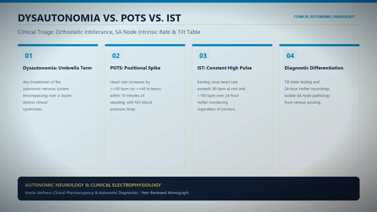
- Postural Orthostatic Tachycardia Syndrome (POTS): Requires a sustained heart rate increment of ≥30 beats per minute (bpm) within 10 minutes of standing (or head-up tilt test) in adults (or ≥40 bpm in individuals aged 12–19), in the absence of orthostatic hypotension (systolic drop >20 mmHg or diastolic drop >10 mmHg), persisting for at least 3 months. When supine, resting heart rate is typically within normal limits (60–80 bpm).
- Inappropriate Sinus Tachycardia (IST): Requires a persistent resting heart rate >90 bpm (with average 24-hour Holter heart rate >90 bpm), and exaggerated tachycardia out of proportion to physiological stressors (such as minimal exertion, emotional arousal, or mild postural shifts). Crucially, the tachycardia is present even when lying flat and during sleep.
Understanding whether heart racing occurs exclusively upon standing or persists regardless of posture is essential. Readers managing autonomic heart rate spikes should also review our clinical analysis of PVCs and PACs in nervous system hyperarousal.
2. Electrophysiology: Sinoatrial Node Autonomy vs. Reflexive Sympathetic Drive
The fundamental divergence between POTS and IST lies in the underlying electrophysiological trigger. The heart's normal pacemaker is the sinoatrial (SA) node, regulated by the balance between sympathetic adrenergic tone and parasympathetic vagal braking (explored in our guide on HRV normative benchmarks and vagal tone).
In Inappropriate Sinus Tachycardia (IST), the pathology is located within the sinoatrial node itself. Studies demonstrate intrinsic SA node hyperactivity characterized by an altered funny current ((I_f)), mediated by hyperpolarization-activated cyclic nucleotide-gated (HCN4) channels. Patients exhibit a blunted sensitivity to acetylcholine and an exaggerated, hyper-responsive chronotropic response to baseline circulating catecholamines.
In POTS, the sinus node is structurally and electrophysiologically normal. The tachycardia is a compensatory neuro-cardiovascular reflex: upon standing, blood pools gravitationally in the lower extremities and splanchnic circulation due to deficient sympathetic vasoconstriction. Venous return to the right atrium plunges, triggering arterial baroreceptors to reflexively fire intense sympathetic catecholamine bursts to maintain cardiac output and cerebral perfusion.
3. Comparative Diagnostic Matrix: 10 Clinical Features
| Clinical Domain | Postural Orthostatic Tachycardia Syndrome (POTS) | Inappropriate Sinus Tachycardia (IST) |
|---|---|---|
| Supine Resting Heart Rate | Normal (typically 60 – 80 bpm) | Elevated (>90 – 100+ bpm) |
| Standing Heart Rate Delta | Sustained ≥30 bpm jump (≥40 in teens) | Variable increase; tachycardia already present supine |
| 24-Hour Holter Average HR | Typically normal (<85 bpm, elevated mainly while upright) | Consistently elevated (>90 bpm over 24 hours) |
| Nocturnal / Sleep Heart Rate | Normalizes into parasympathetic bradycardia (50–65 bpm) | Often remains persistently elevated (75–90+ bpm) |
| Primary Mechanism | Reflexive compensation for venous pooling and hypovolemia | Intrinsic sinoatrial node dysfunction / enhanced (I_f) current |
| Peripheral Blood Pooling | Prominent acrocyanosis, mottled red/purple legs on standing | Absent or minimal; microvascular tone generally intact |
| Brain Fog on Standing | Severe cerebral hypoperfusion (15–20% CBF velocity drop) | Mild to moderate; less dependent on orthostatic posture |
| Response to Saline Infusion | Dramatic heart rate reduction and symptom relief | Minimal or negligible effect on basal tachycardia |
| First-Line Pharmacotherapy | Fludrocortisone, Midodrine, Pyridostigmine, Propranolol | Ivabradine (Corlanor), Beta-Blockers |
| Non-Pharmacological Staples | Waist-high compression, 3–10g sodium, 3L fluid intake | Graded aerobic training, stress reduction, vagal exercises |
4. Testing Workup: Tilt Table, 24-Hour Holter & Autonomic Reflex Screen
A rigorous diagnostic workup is paramount to avoid labeling genuine dysautonomia as "psychogenic panic" or anxiety. The clinical evaluation should incorporate:
- 12-Lead Resting ECG: Confirms normal sinus P-wave morphology (ruling out ectopic atrial tachycardia, atrial flutter, or AV nodal reentrant tachycardia). In IST, the P wave is identical to sinus rhythm.
- 24-Hour to 14-Day Ambulatory Holter Monitor: Assesses circadian heart rate dynamics. IST is confirmed when average 24-hour heart rate exceeds 90 bpm, with sleeping rates remaining inappropriately accelerated. In POTS, the Holter reveals abrupt spikes corresponding with waking upright activity and complete normalization during sleep.
- Head-Up Tilt Table Test (HUTT) or NASA 10-Minute Lean Test: Patients are evaluated supine for 10 minutes, followed by passive 70-degree head-up tilt. Blood pressure, continuous beat-to-beat ECG, and cerebral oximetry are recorded. This differentiates POTS from orthostatic hypotension and psychogenic pseudosyncope.
- Comprehensive Metabolic and Endocrine Screen: TSH, free T4, 24-hour urine metanephrines (to exclude pheochromocytoma), and baseline morning cortisol (see our guide on cortisol reference charts by time of day).
5. Overlapping Syndromes and Autoantibody Biomarkers
Emerging research from the Mayo Clinic and Vanderbilt Autonomic Dysfunction Center has identified common autoimmune pathophysiologies connecting POTS and IST. Approximately 15% of patients meet criteria for both conditions simultaneously—a state termed POTS-IST overlap.
High titers of functional G-protein coupled receptor (GPCR) autoantibodies—specifically against β1-adrenergic, β2-adrenergic, and muscarinic M2 acetylcholine receptors—have been documented in up to 60% of post-viral dysautonomia cohorts. When autoantibodies stimulate β1 receptors, intrinsic sinus tachycardia occurs (IST phenotype); when autoantibodies block peripheral α1-adrenergic receptors, vasoconstrictive failure causes postural venous pooling (POTS phenotype).
6. Therapeutic Management: Ivabradine, Beta-Blockers & Volume Expansion
Because the physiological mechanisms differ, pharmacological regimens must be customized:
- Ivabradine (HCN Channel Blocker): The undisputed drug of choice for Inappropriate Sinus Tachycardia. By selectively inhibiting the cardiac pacemaker (I_f) current in the sinus node, ivabradine slows heart rate without lowering blood pressure or depressing cardiac contractility, preserving exercise tolerance. It is also increasingly utilized off-label in hyperadrenergic POTS.
- Volume Expansion Protocol: For POTS, expanding intravascular volume with 3 to 5 grams of dietary sodium and 3 liters of water daily is fundamental. Fludrocortisone (0.1 to 0.2 mg daily) promotes renal sodium retention to prevent orthostatic drops.
- Peripheral Vasoconstrictors (Midodrine): An α1-adrenergic agonist that constricts venous capacitance beds, preventing blood pooling in the lower extremities during standing. Midodrine is ineffective in isolated IST.
- Parasympathetic Enhancers (Pyridostigmine): An acetylcholinesterase inhibitor that prolongs acetylcholine availability at autonomic ganglia, enhancing vagal braking and improving standing hemodynamic stability (learn more about exercises to raise vagal tone).