
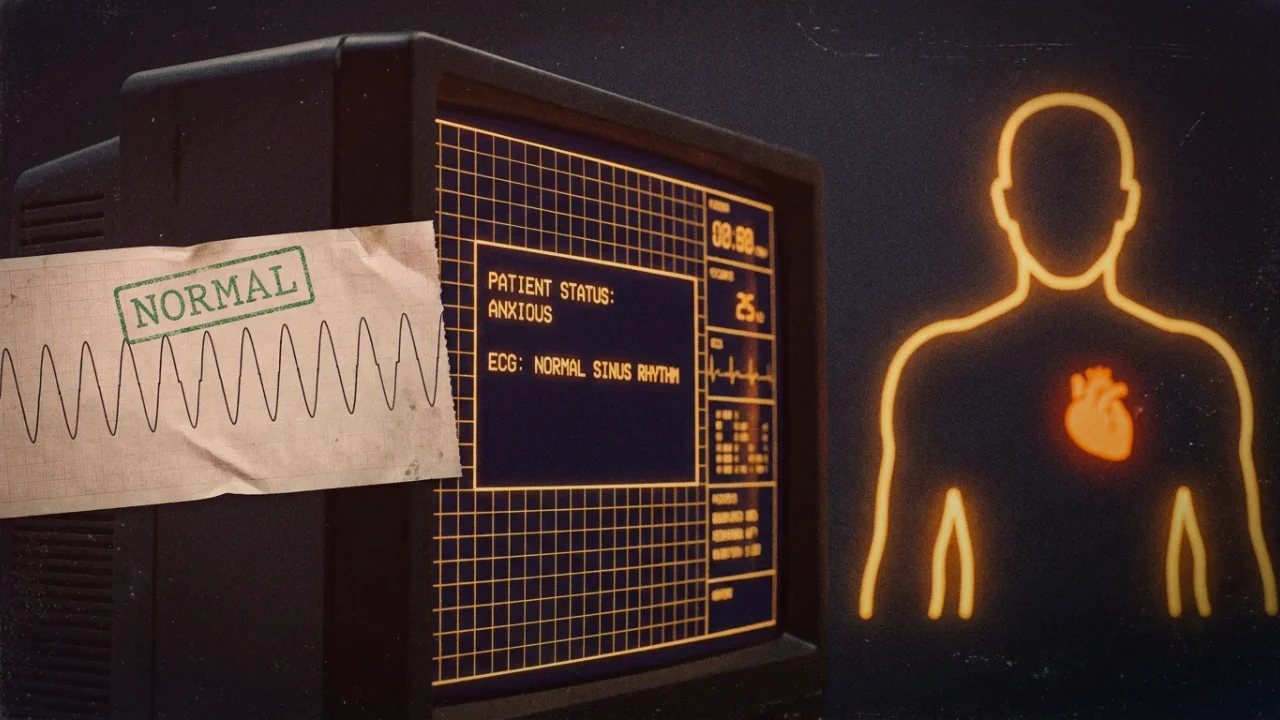
1. What Are PVCs and PACs? The Cardiac Conduction System
In a healthy heart, electrical depolarization begins in the sinoatrial (SA) node—the physiological pacemaker located in the upper right atrium. The electrical wave propagates smoothly across the atria, pauses momentarily at the atrioventricular (AV) node to allow ventricular filling, and rapidly travels down the Bundle of His and Purkinje fibers to trigger coordinated ventricular systole.
Premature beats represent electrical insubordination. An irritable cell or cluster of cells outside the SA node—known as an ectopic focus—spontaneously depolarizes before the SA node has fired its scheduled impulse. If this ectopic discharge occurs in the atria, it is a Premature Atrial Contraction (PAC); if it originates in the Purkinje network or ventricular myocardium, it is a Premature Ventricular Contraction (PVC).
2. The Anatomy of the "Thud": The Compensatory Pause Explained
Most patients believe the alarming sensation is the premature beat itself. In reality, the premature beat occurs so early in the cardiac cycle that the ventricles have had almost no time to fill with blood from the atria. Consequently, the premature contraction generates negligible stroke volume, producing a pulse wave so weak that it often cannot be felt at the radial wrist artery.
Immediately following the premature depolarization, the ventricular myocardium enters an absolute refractory period. When the SA node fires its next normal electrical signal, the ventricular muscle is electrically unreceptive and cannot contract. The heart must wait for the subsequent regular SA node cycle, creating a prolonged silence known as the full compensatory pause.
During this extra-long pause, blood continues to pool into the relaxed ventricles from the venous circulation, stretching the myocardial fibers to capacity. When the next normal sinus impulse arrives, Starling’s Law of the Heart takes effect: the overfilled ventricles contract with immense mechanical force, slamming the heart against the anterior chest wall. That violent "thump," "flip," or "drop" is the forceful post-pause beat, not the missed beat.
Cardiology Fact: Ectopic Burden vs. Clinical Risk
In 24-hour Holter monitoring studies, having between 10 and 100 isolated PVCs per day is documented in over 70% of completely healthy adult humans. Cardiologists generally consider PVCs benign and hemodynamically inconsequential unless the total 24-hour ectopic burden exceeds 10% to 15% of total heartbeats (roughly 10,000 to 15,000 PVCs daily) or co-occurs with structural heart disease.
3. The Adrenaline Cascade: Calcium Overload in Ectopic Foci
Why does psychological stress provoke these ectopic discharges? Under acute anxiety or chronic hypervigilance, the sympathetic nervous system and adrenal medulla flood the coronary circulation with epinephrine and norepinephrine.
These catecholamines bind avidly to beta-1 adrenergic receptors on cardiomyocytes. Receptor activation stimulates adenylyl cyclase, boosting intracellular cyclic AMP (cAMP) and activating Protein Kinase A (PKA). PKA phosphorylates L-type calcium channels and the ryanodine receptor-2 (RyR2) on the sarcoplasmic reticulum. Intracellular calcium surges into the cytosol. In sensitive myocardial cells, this excess calcium triggers delayed afterdepolarizations (DADs)—sub-threshold membrane oscillations that reach the voltage gate and fire an unprovoked premature electrical wave.
4. Why Anxiety Causes Heart Palpitations and Skipped Beats
The relationship between stress and cardiac ectopy is a classic reciprocal loop. Understanding why anxiety causes heart palpitations and skipped beats is essential to breaking the vicious psychological cycle that keeps ectopic foci firing.
- Sensory Interoceptive Magnification: Anxiety sensitizes the anterior insular cortex. Healthy people experience isolated PVCs without noticing them; anxious individuals feel every single ventricular contraction with terrifying clarity.
- Hyperventilation & Hypocapnia: Anxious shallow breathing blows off carbon dioxide, causing mild respiratory alkalosis. Alkalosis reduces ionized serum calcium and magnesium, directly destabilizing myocardial resting membrane potentials.
- Vagal Withdrawal: Chronic stress removes the cholinergic parasympathetic brake. Acetylcholine released by the vagus nerve normally hyperpolarizes pacemaker cells and suppresses ectopic automaticity; its absence invites ventricular irritability.
5. Benign Ectopy vs. Structural Arrhythmia: Clinical Red Flags
While the overwhelming majority of palpitations in young and middle-aged adults are benign stress-induced PVCs/PACs, medical evaluation is non-negotiable to establish structural safety. The following clinical matrix delineates benign ectopy from serious cardiac pathology:
| Clinical Parameter | Benign Anxiety PVCs / PACs | Pathological / Red Flag Arrhythmia |
|---|---|---|
| Timing of Onset | During rest, lying in bed, or post-stress downtime | Onset at peak physical exercise or exertion |
| Associated Symptoms | Transient startle, nervous throat catch, anxiety | True syncope (fainting), chest pressure, diaphoresis |
| ECG Morphology | Isolated, monomorphic QRS, normal sinus background | Polymorphic PVCs, R-on-T phenomenon, runs of VT |
| Structural Heart Status | Normal echocardiogram (LVEF > 55%), no hypertrophy | Ischemic scar, cardiomyopathy, valvular stenosis |
6. Electrophysiological Reset: Calming Premature Beats Naturally
For individuals cleared of structural cardiac disease by a cardiologist, calming ectopic activity requires targeted stabilization of cardiomyocyte resting potentials and vagal reinstatement:
- Magnesium Glycinate or Taurate Repletion: Magnesium acts as a natural physiological calcium channel blocker, regulating the sodium-potassium ATPase pump and suppressing delayed afterdepolarizations in myocardial tissue.
- Valsalva Maneuver / Modified Carotid Pacing: Exhaling forcefully against closed airways for 10 seconds stimulates aortic baroreceptors, triggering immediate vagal acetylcholine release directly onto cardiac muscarinic-2 receptors.
- Electrolyte Osmoregulation: Ensuring adequate sodium and potassium intake to maintain myocardial transmembrane electrical gradients; dehydration-induced hypokalemia is a primary catalyst for PVC clusters.
- Postural Decoupling: If PVCs trigger predominantly when reclining, transitioning to the right lateral decubitus position minimizes anatomical cardiac impingement against the anterior thoracic cage.