
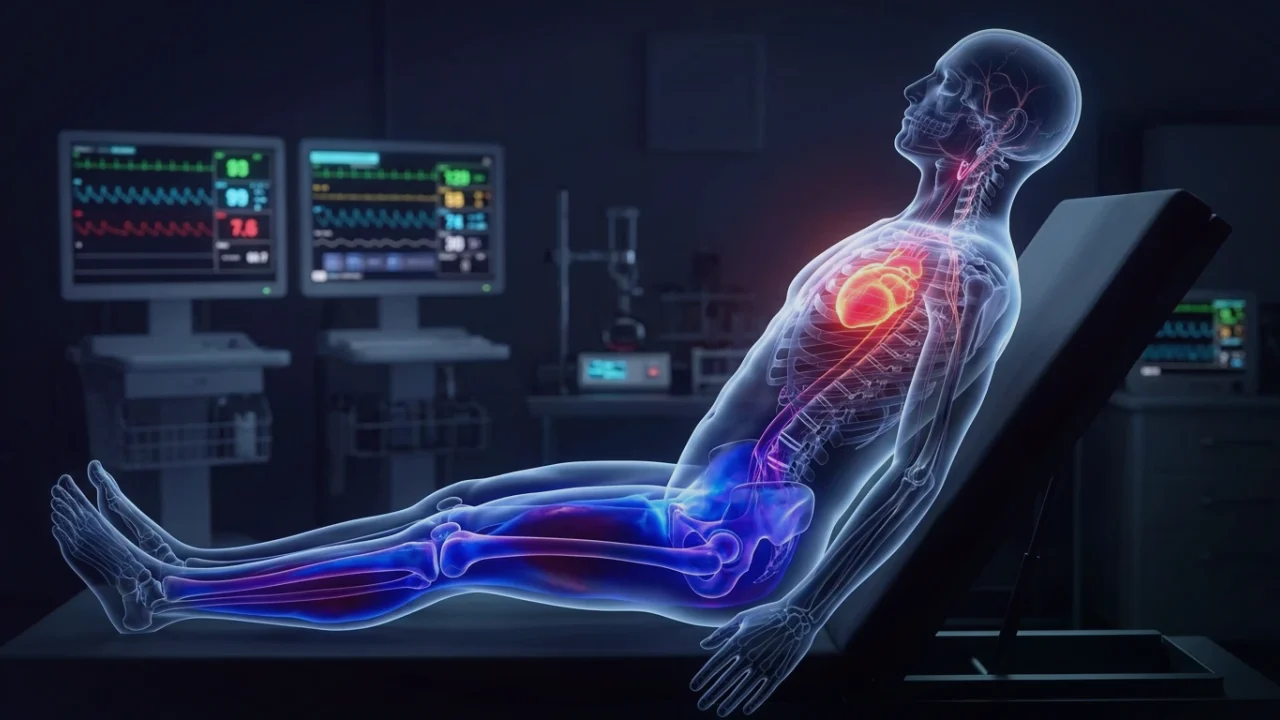
1. Hydrostatic Pressure and the Skeletal Muscle Pump
In a standing adult, gravity exerts immense hydrostatic pressure on the vascular tree. For every centimeter below the right atrium, intravascular venous pressure increases by roughly 0.77 mmHg, reaching approximately 90 to 100 mmHg in the feet. Under normal physiological conditions, the body counteracts this gravitational pull through two mechanisms: rhythmic contraction of the calf skeletal muscle pump and active sympathetic vasoconstriction mediated by α1-adrenergic receptors on venous capacitance vessels.
In patients with autonomic neuropathy, Postural Orthostatic Tachycardia Syndrome (POTS), and Ehlers-Danlos Syndrome (EDS), this vasoconstrictive reflex fails. The collagen matrix of the vein walls is either excessively compliant or sympathetic denervation prevents smooth muscle tone from stiffening. Up to 800 mL of blood accumulates rapidly in dependent vascular beds within minutes of standing.
This failure of venous return is closely linked to somatic panic and heart-rate spikes, as detailed in our guide on panic attacks vs heart attack symptoms.
2. The Hidden Culprit: Mesenteric Splanchnic Capacitance
Most patients focus on the visible swelling and reddish-purple discoloration in their feet and calves. However, cardiovascular physiology reveals that the splanchnic-mesenteric circulation—the massive network of veins supplying the stomach, intestines, liver, and spleen—serves as the primary blood reservoir of the human body, holding up to 25% to 30% of total blood volume at rest.
Because the splanchnic vascular bed is compliant and highly innervated by the greater splanchnic sympathetic nerves, sympathetic failure results in massive mesenteric venous pooling. Blood becomes sequestered in the gut, reducing venous return through the inferior vena cava into the right ventricle, starving stroke volume, and depriving the brain of essential blood flow.
3. Clinical Presentation: Dependent Acrocyanosis & Edema
Clinically, peripheral venous pooling presents as dependent acrocyanosis: within 3 to 5 minutes of standing or sitting with legs hanging down, the lower extremities turn mottled red, dark purple, or bluish. Patients report sensations of intense heaviness, pulsating fullness, throbbing pain, and burning heat in the feet.
When hydrostatic pressure exceeds oncotic capillary pressure, plasma fluid extravasates across the endothelial barrier into interstitial tissues, causing orthostatic dependent edema. The resulting loss of intravascular circulating volume triggers the renin-angiotensin-aldosterone axis to retain water, often accompanied by paradoxical morning facial puffiness.
4. The Medical Compression Hierarchy: Abdominal vs. Waist vs. Knee
Clinical trials conducted by the Vanderbilt Autonomic Dysfunction Center evaluated the hemodynamic impact of various compression garments in POTS patients using tilt table testing. The findings fundamentally altered clinical practice guidelines:
| Compression Modality | Pressure Rating | Impact on Upright Heart Rate | Impact on Stroke Volume | Clinical Verdict |
|---|---|---|---|---|
| Knee-High Socks | 15 – 20 mmHg | Negligible (<3 bpm reduction) | Minimal change (<5%) | Ineffective as monotherapy for POTS |
| Thigh-High Stockings | 20 – 30 mmHg | Mild reduction (5 – 8 bpm) | Moderate improvement (+10%) | Helpful, but ignores splanchnic pooling |
| Abdominal Binder Alone | 20 – 30 mmHg | Significant reduction (10 – 14 bpm) | Strong increase (+18%) | Highly Effective; compresses mesenteric reservoir |
| Waist-High Tights + Abdominal Binder | 30 – 40 mmHg | Maximal reduction (15 – 22 bpm) | Maximal restoration (+26%) | Gold Standard clinical intervention |
The clinical takeaway is decisive: compression must cover the abdomen. Compressing the calves alone leaves the massive splanchnic reservoir untouched. Patients should wear medical-grade class II (20–30 mmHg) or class III (30–40 mmHg) waist-high compression tights, or pair thigh-high stockings with a wide elastic abdominal binder.
5. Vagal Cardiopulmonary Reflexes and Atrial Stretch Receptors
The vagus nerve is intimately coupled to venous return via low-pressure cardiopulmonary mechanoreceptors (Type A and Type B atrial receptors) located at the veno-atrial junctions. When venous return is adequate, the cardiac atria stretch during diastole, stimulating unmyelinated vagal afferents in the Nucleus Tractus Solitarius (NTS) to sustain resting parasympathetic tone.
When chronic blood pooling deprives the atria of diastolic stretch, these vagal afferents fall silent. The brainstem misinterprets the empty cardiac chambers as acute hemorrhagic hypovolemia, unleashing uninhibited sympathetic tachycardia. Restoring venous return via medical compression restores atrial stretch, re-engaging the cardiac vagal brake (explore how to restore vagal signaling in our clinical vagal tone protocol).