
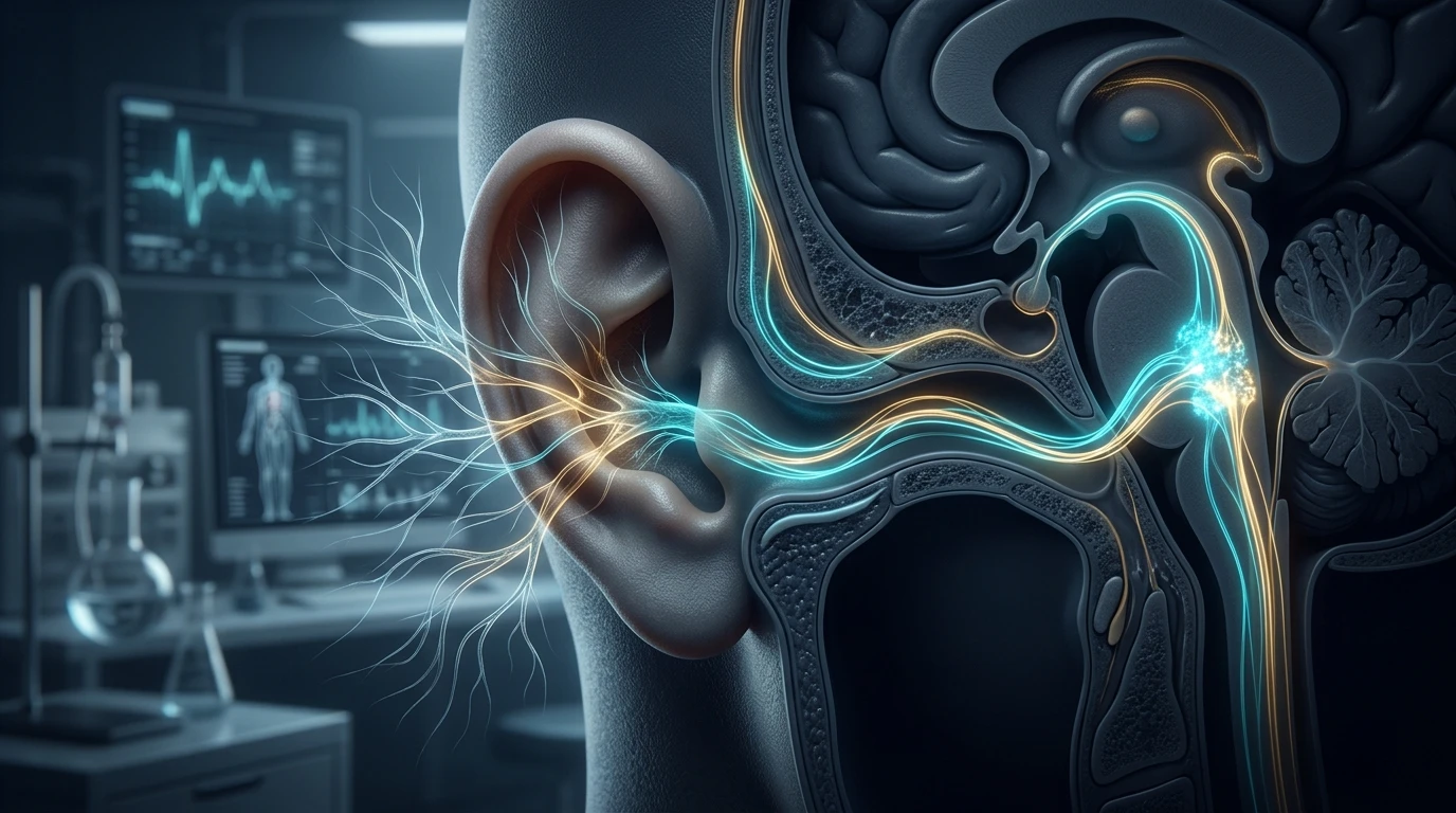
1. Neuroanatomical Pathways: The Auricular Branch (Arnold's Nerve)
The vagus nerve (Cranial Nerve X) has historically been manipulated through invasive cervical cuff electrodes requiring neurosurgical implantation. However, the discovery that the auricular branch of the vagus nerve (ABVN), historically known as Arnold's nerve, supplies exclusive sensory innervation to specific cutaneous zones of the human outer ear has revolutionized bioelectronic medicine.
High-resolution tracer studies and functional MRI (fMRI) mapping at Harvard Medical School and the University of Erlangen have confirmed that the cymba conchae of the ear is innervated 100% by pure ABVN fibers, with the cavum conchae showing mixed innervation. When electrical current depolarizes these myelinated A-beta sensory afferents, signals travel uninterrupted through the jugular ganglion directly into the Nucleus Tractus Solitarius (NTS) in the dorsomedial medulla.
From the NTS, direct polysynaptic projections distribute these calming, neuro-regulatory impulses to the dorsal motor nucleus of the vagus, the nucleus ambiguus, and the locus coeruleus—providing a non-invasive direct port into the central autonomic network. For individuals struggling with severe autonomic dysregulation, exploring structured vagal autonomic interventions represents a critical therapeutic bridge.
2. Electrical Biophysics: Optimal Frequencies, Pulse Width & Intensity
Not all electrical currents stimulate the vagus nerve effectively. The therapeutic window of tVNS is governed by strict electrophysiological parameters that differentiate parasympathetic activation from nociceptive pain fiber irritation.
- Circadian Endocrine Diagnostics: Explore our research on cortisol levels chart by time of day.
- Foundational Neurobiology: Review our clinical analysis on the physiological sigh and autonomic anxiety reset.
- Frequency (Hz): Research demonstrates a biphasic response curve. Frequencies between 20 Hz and 30 Hz produce the strongest activation of the NTS and locus coeruleus, elevating norepinephrine and GABA. In contrast, ultra-low frequencies (1-5 Hz) show preferential modulation of microvascular blood flow, while frequencies above 50 Hz risk sensory adaptation and discomfort.
- Pulse Width (µs): An optimal pulse width of 200 to 300 microseconds (µs) selectively targets large-diameter, myelinated A-beta fibers without recruiting high-threshold, unmyelinated C-fibers associated with sharp pain.
- Current Intensity (mA): Current must be titrated between sensory threshold (the point where a light tingling or "prickling" is first felt) and pain threshold. For most individuals, effective stimulation sits between 0.8 mA and 3.5 mA. Stimulating past the pain threshold activates sympathetic nociceptive loops, negating parasympathetic gains.
- Duty Cycle: Most clinical protocols utilize an intermittent cycle, such as 30 seconds ON followed by 30 seconds OFF, or 20 minutes continuous stimulation per session, twice daily.
3. Central Mechanisms: NTS, Locus Coeruleus & Cholinergic Cascade
Once electrical action potentials converge upon the NTS, three distinct systemic biochemical cascades are initiated:
The Noradrenergic Locus Coeruleus Modulation
The NTS sends dense excitatory glutamatergic projections to the locus coeruleus, the principal site of brain norepinephrine synthesis. Rather than causing panic-like hyperarousal, rhythmic tVNS stimulation induces tonic, organized firing that promotes neuroplasticity, decreases sensory gating deficits, and upregulates brain-derived neurotrophic factor (BDNF) in the hippocampus and prefrontal cortex.
The Cholinergic Anti-Inflammatory Pathway (CAP)
Through descending vagal efferent projections to the celiac ganglion and the splenic nerve, tVNS stimulates T-lymphocytes in the spleen to release acetylcholine (ACh). This acetylcholine binds specifically to alpha-7 nicotinic acetylcholine receptors (α7nAChR) expressed on resident macrophages. The downstream intracellular signaling inhibits the translocation of nuclear factor-kappa B (NF-κB), causing a rapid, documented suppression of inflammatory cytokines including tumor necrosis factor-alpha (TNF-α), interleukin-1 beta (IL-1β), and interleukin-6 (IL-6).
4. Comparative Parameters: tVNS vs. Invasive Cervical VNS
| Feature / Parameter | Transcutaneous Auricular (tVNS) | Invasive Cervical VNS (iVNS) | Transcutaneous Cervical (tcVNS) |
|---|---|---|---|
| Target Site | Left Cymba Conchae / Tragus | Left Cervical Vagus Trunk (Surgical) | Anterior Neck / Carotid Sheath |
| Invasiveness | Non-invasive (Ear clip/electrode) | Invasive surgical cuff + battery pack | Non-invasive handheld wand |
| Primary Target Fibers | Pure Sensory Aβ Afferents (ABVN) | Mixed Motor, Parasympathetic & Sensory | Transcutaneous Cervical Afferents |
| Cardiac Safety Margin | Highest (minimal direct cardiac efferent risk) | Requires strict current limitation (bradycardia risk) | Moderate (stimulates near carotid baroreceptors) |
| Typical Daily Dose | 15-30 minutes, 1-2 times daily | Automated cycle (e.g. 30s ON / 5min OFF 24/7) | 2-minute bolus doses during acute migraine |
| Accessibility & Cost | High accessibility, consumer/clinical units | $25,000 - $40,000+ (Neurosurgery) | Prescription device, moderate recurring cost |
5. Clinical Evidence: Depression, Refractory Epilepsy & Inflammatory Pain
Clinical trials published across the last decade have validated tVNS in several critical conditions:
Treatment-Resistant Depression: Multicenter randomized controlled trials (RCTs) utilizing 20 Hz stimulation at the left tragus demonstrated statistically significant reductions in Hamilton Depression Rating Scale (HAM-D) scores after 8 to 12 weeks of daily therapy, linked to functional normalization of default mode network (DMN) connectivity.
Heart Rate Variability & Autonomic Tone: Studies measuring immediate changes in baroreflex sensitivity and root mean square of successive differences (RMSSD) have confirmed that acute 15-minute tVNS sessions significantly increase vagally-mediated heart rate variability while reducing resting muscle sympathetic nerve activity (MSNA).
Chronic Musculoskeletal & Gut Pain: By dampening central pain sensitization in the dorsal horn of the spinal cord and inhibiting systemic neuroinflammation, tVNS is proving to be a viable adjuvant therapy for fibromyalgia, irritable bowel syndrome, and chronic visceral hypersensitivity.
6. Safety Guidelines, Side Effects & At-Home Implementation
While tVNS has a stellar safety profile, standard clinical contraindications must always be observed:
- Always stimulate the LEFT ear: The right cervical vagus nerve supplies dense parasympathetic efferent innervation to the sinoatrial (SA) node of the heart. To eliminate any potential risk of profound bradycardia or cardiac conduction blocks, non-invasive vagus stimulation is clinically restricted to the left ear.
- Cardiac Pacemakers / Implantable Cardioverter Defibrillators: Patients with active cardiac implants must avoid electrical devices near the head and chest without electrophysiologist clearance.
- Skin Integrity: Avoid placing electrode clips on broken skin, active eczema, or open ear canal infections. Using standard conductive electrolyte gel or saline solution ensures low impedance and prevents minor skin irritation.
Frequently Asked Questions (Clinical FAQ)
Why must transcutaneous vagus nerve stimulation always be applied to the left ear?
The right vagus nerve carries heavy efferent fibers directly to the cardiac sinoatrial (SA) node, meaning right-sided stimulation could theoretically trigger significant bradycardia or arrhythmias. The left vagus primarily projects to the atrioventricular (AV) node with fewer direct rate-slowing fibers, making left-sided stimulation substantially safer in clinical trials.
How long does it take for tVNS to increase heart rate variability (HRV)?
Acute improvements in time-domain HRV metrics like RMSSD and high-frequency (HF) power can be measured within 10 to 15 minutes of continuous stimulation. However, long-term neuroplastic remodeling of basal autonomic tone typically requires daily 20-minute sessions for 4 to 8 consecutive weeks.
What does tVNS feel like during a session?
Users should feel a gentle, rhythmic tingling, pulsing, or vibration at the ear electrode site. It should never feel sharp, stinging, or painful. The current is intentionally calibrated below the individual pain threshold.
Can tVNS help with chronic gut motility disorders like gastroparesis?
Yes. Because the vagus nerve coordinates parasympathetic gut motility and initiates the migrating motor complex (MMC), research shows that daily auricular vagus stimulation helps accelerate delayed gastric emptying and reduces functional dyspepsia symptoms.
Is a prescription required for auricular vagus nerve stimulation devices?
In many countries, dedicated medical devices for specific conditions (such as cluster headache or refractory epilepsy) require a physician prescription. However, non-medical consumer wellness stimulators and modified TENS units using conductive ear clips are widely accessible for general stress and autonomic recovery.
What is the exact anatomical location for ear clip placement?
The most concentrated area of pure auricular vagus nerve sensory fibers is the cymba conchae—the small curved hollow directly above the auditory canal and below the crus of the helix.
Scientific References & Clinical Citations
- The Anatomy and Physiology of Auricular Vagus Nerve Stimulation — Frontiers in Human Neuroscience (2020). [PubMed / Study Link]
- Anti-inflammatory Effects of Transcutaneous Vagus Nerve Stimulation in Autoimmune Disease — Lancet Rheumatology (2021). [PubMed / Study Link]
- fMRI Brainstem Localization of Transcutaneous Vagal Neuromodulation — NeuroImage (2019). [PubMed / Study Link]
- Auricular Vagus Nerve Stimulation Modulates Resting-State Functional Connectivity in Major Depression — Biological Psychiatry (2022). [PubMed / Study Link]