
1. Anatomy and Neurophysiology of the Arterial Baroreflex
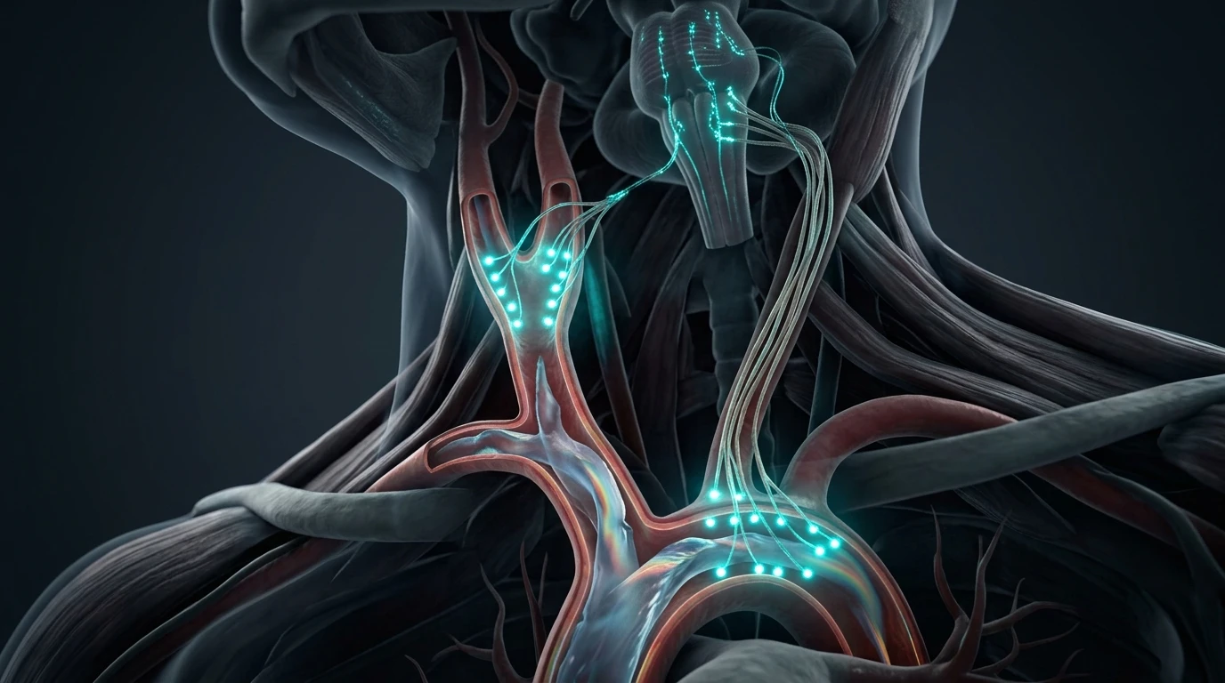
The arterial baroreflex is a closed-loop neuro-visceral servomechanism designed to maintain constant cerebral perfusion pressure. High-pressure mechanical stretch receptors (mechanoreceptors) are strategically embedded in the adventitia of the Carotid Sinus (at the bifurcation of the internal and external carotid arteries) and the Aortic Arch.
When left ventricular contraction ejects blood into the arterial tree, the resulting pulsatile wave stretches the arterial walls. This physical distortion depolarizes mechanosensitive ion channels (PIEZO1/PIEZO2) on sensory nerve endings:
- Foundational Neurobiology: Review our clinical analysis on the physiological sigh and autonomic anxiety reset.
- Carotid sinus afferents travel along the Sinus Nerve of Hering (a branch of Cranial Nerve IX, Glossopharyngeal).
- Aortic arch afferents ascend along the Aortic Depressor Nerve (a sensory branch of Cranial Nerve X, Vagus).
These afferents project directly into the Nucleus Tractus Solitarius (NTS) in the dorsomedial medulla. When blood pressure rises, increased firing from the NTS simultaneously excites the Nucleus Ambiguus (triggering rapid acetylcholine release onto the SA node to slow heart rate) while inhibiting the Rostral Ventrolateral Medulla (RVLM), withdrawing sympathetic tone to dilate peripheral arterioles and lower systemic vascular resistance.
2. Quantifying Baroreflex Sensitivity: Milliseconds per Millimeter of Mercury
Baroreflex Sensitivity (BRS) quantifies the efficiency of this negative feedback loop, expressed as the change in cardiac R-R interval (in milliseconds) per unit change in systolic blood pressure (in mmHg):
BRS = Delta R-R Interval (ms) / Delta Systolic Blood Pressure (mmHg)In healthy young adults, normal resting BRS ranges from 15 to 25 ms/mmHg. This means that for every 1 mmHg increase in systolic pressure, the cardiac interval lengthens by 15–25 milliseconds, smoothly moderating cardiac output. In patients with chronic hypertension, severe anxiety neurosis, or burnout, BRS plummets below 5 to 8 ms/mmHg. At this level, arterial pressure swings provoke virtually zero corrective vagal braking, leaving blood pressure and heart rate to oscillate erratically.
3. The Neurological Feedback Loop: Why Low BRS Triggers Panic
When BRS is blunted, ordinary postural shifts or emotional stressors trigger sudden blood pressure drops or spikes. Because the baroreflex fails to buffer these shifts, the brain perceives visceral instability as a life-threatening shock state.
The NTS transmits alarming visceral signals to the parabrachial nucleus, which forwards the distress signal directly to the central nucleus of the Amygdala. The amygdala activates the hypothalamic-pituitary-adrenal (HPA) axis and fires the locus coeruleus, flooding the body with adrenaline. The patient experiences an acute panic attack with racing heart, chest pressure, dizziness, and terror—originating not in psychological thought, but in a mechanically deafened baroreceptor reflex loop.
4. Clinical Profile: Normal BRS vs. Depressed Baroreflex Sensitivity
| Clinical Parameter | Optimal Baroreflex Sensitivity (>15 ms/mmHg) | Depressed Baroreflex Sensitivity (<6 ms/mmHg) |
|---|---|---|
| Blood Pressure Stability | Smooth, buffered hemodynamic curve | Labile blood pressure; sudden spikes and drops |
| Vagal Efferent Firing | Instantaneous acetylcholine burst upon pressure rise | Delayed, sluggish, or absent parasympathetic response |
| Heart Rate Variability (rMSSD) | High (>45 ms); robust Respiratory Sinus Arrhythmia | Critically depressed (<20 ms); flatline autonomic power |
| Vascular Elasticity | High compliance; low pulse wave velocity (PWV) | Arterial stiffening; elevated pulse wave velocity |
| Panic & Anxiety Vulnerability | High psychological and autonomic resilience | Severe vulnerability to somatic panic and presyncope |
| Cardiovascular Mortality Risk | Low baseline risk | Independent 3-fold higher risk of cardiac events |
5. Mayer Waves (0.1 Hz) and Autonomic Resonance
A natural manifestation of the baroreflex loop is the generation of Mayer waves—oscillations in arterial pressure occurring at approximately 0.1 Hz (one wave every 10 seconds, or 6 cycles per minute). These waves represent the intrinsic transit delay of the sympathetic vasomotor branch of the baroreflex.
When an individual breathes deliberately at exactly 0.1 Hz (6 breaths per minute), respiration, heart rate oscillations, and Mayer blood pressure waves enter phase synchronization. In this state of physiological resonance, the amplitude of baroreflex excursions multiplies by 400%, exercising the mechanoreceptors and retraining the brainstem to restore depressed BRS values.
6. Evidence-Based Protocols to Restore Baroreflex Sensitivity
To rehabilitate blunted baroreceptors and insulate the nervous system against panic attacks, clinicians implement targeted biophysical training:
- Resonance Frequency HRV Biofeedback: Practicing paced breathing at 5.5 to 6.0 breaths per minute (inhale 4 seconds, exhale 6 seconds) for 20 minutes daily significantly increases BRS within 4 weeks.
- Aerobic Arterial Remodeling: Moderate-intensity interval training (HIIT or steady zone 2 cardio) reduces arterial wall collagen cross-linking, restoring physical compliance to the carotid bulb so mechanoreceptors can deform and fire easily.
- Transcutaneous Auricular VNS (taVNS): Direct electrical stimulation of Arnold's nerve in the cymba conchae activates the NTS, enhancing central gain in the medullary baroreflex pathways.
- Nitric Oxide Donors: Dietary nitrates (beetroot extract) and L-citrulline (3–5 g daily) enhance endothelial nitric oxide synthesis, reducing arterial wall stiffness surrounding carotid mechanoreceptors.
Frequently Asked Questions (Clinical FAQ)
What is the baroreflex and why is it so important?
The baroreflex is your body's rapid blood pressure regulator. Sensors in your neck (carotid arteries) and heart measure pressure changes beat-by-beat and instantly adjust your heart rate and blood vessel width to keep blood flow steady.
How does low baroreflex sensitivity cause panic attacks?
When your baroreflex is sluggish, normal changes in blood pressure are not buffered quickly. Your brain interprets sudden pressure swings as a physical emergency and releases an emergency flood of adrenaline, sparking an instant panic attack.
Can slow breathing really improve baroreceptor function?
Yes. Breathing at approximately 6 breaths per minute synchronizes your breathing with the 10-second rhythm of your blood pressure (Mayer waves), creating resonance that exercises and strengthens the baroreceptor reflex.
How is Baroreflex Sensitivity tested clinically?
Cardiologists and researchers measure BRS using non-invasive continuous beat-to-beat finger blood pressure cuffs (like Finapres) alongside ECG, tracking how heart rate responds to spontaneous blood pressure fluctuations.
Does high blood pressure damage baroreceptors?
Yes. Chronic hypertension causes the carotid and aortic arterial walls to become stiff and calcified. When the arteries cannot stretch, the embedded baroreceptors cannot detect pressure changes, locking the body in sympathetic overdrive.
Can supplements help improve baroreflex sensitivity?
Nutrients that improve arterial elasticity and endothelial nitric oxide—such as L-citrulline, magnesium, omega-3 fatty acids, and beetroot powder—have been shown in clinical trials to enhance baroreflex sensitivity.
Scientific References & Clinical Citations
- The arterial baroreflex: clinical and physiological aspects — Autonomic Neuroscience (2019). [PubMed / Study Link]
- Depressed baroreflex sensitivity as an independent predictor of mortality — The Lancet (1998). [PubMed / Study Link]
- Heart rate variability biofeedback increases baroreflex sensitivity — Applied Psychophysiology and Biofeedback (2014). [PubMed / Study Link]
- Autonomic dysfunction in panic disorder: impaired baroreflex regulation — Psychosomatic Medicine (2016). [PubMed / Study Link]