
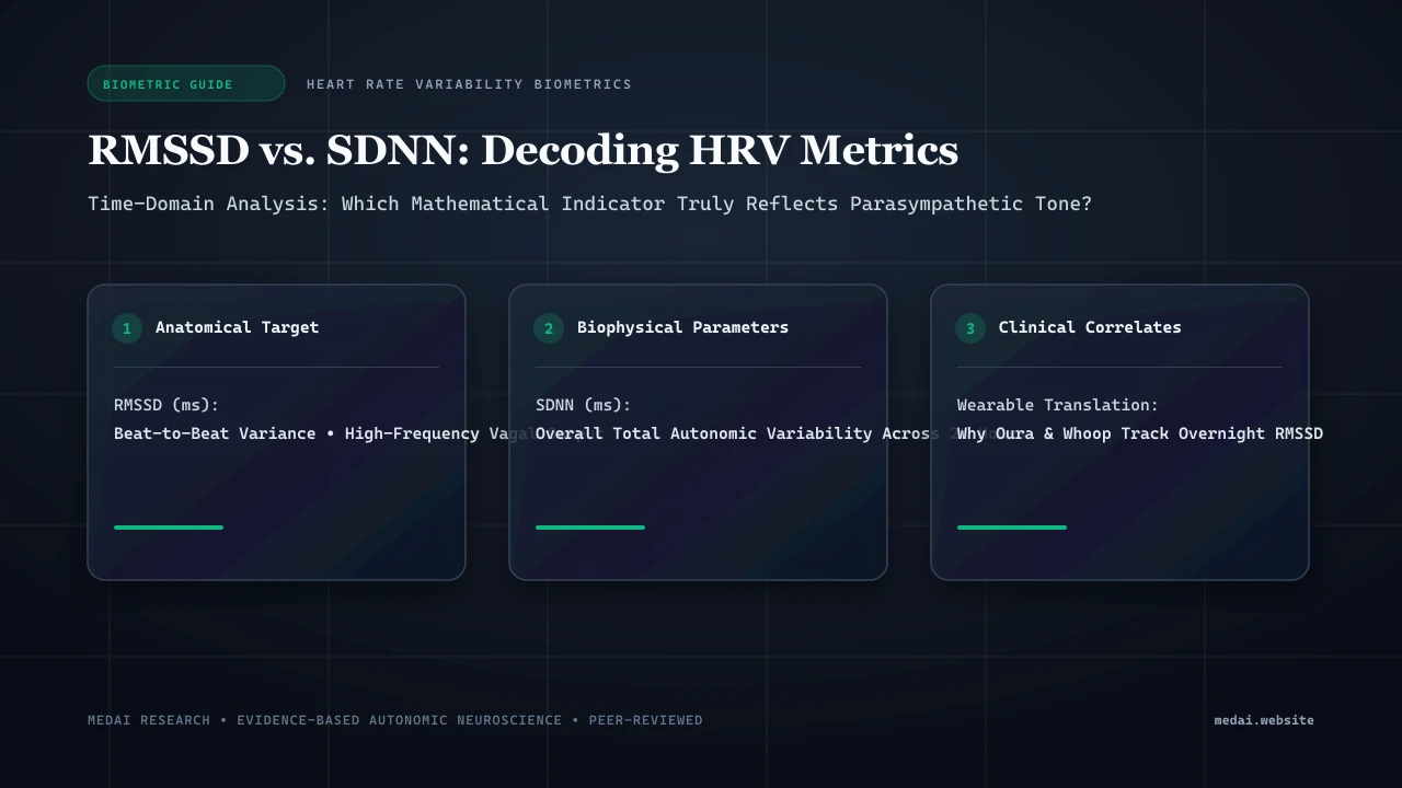
1. Mathematical Anatomy: How rMSSD and SDNN Are Calculated
Every heartbeat is initiated by the sinoatrial (SA) node, but the interval between beats—the R-R or normal-to-normal (NN) interval—is continuously varied by autonomic inputs. Time-domain mathematical algorithms convert these millisecond variations into actionable biomarkers:
rMSSD (Root Mean Square of Successive Differences)
The mathematical formulation is: rMSSD = sqrt( (1 / (N - 1)) * SUM[ (RR_(i+1) - RR_i)^2 ] ). By calculating differences exclusively between consecutive beats, squaring them, and taking the root mean square, rMSSD acts as a pure high-pass filter. Slow circadian shifts or postural drifts are mathematically eliminated, isolating rapid millisecond adjustments.
SDNN (Standard Deviation of NN Intervals)
The formulation is: SDNN = sqrt( (1 / (N - 1)) * SUM[ (RR_i - Mean_RR)^2 ] ). Here, every interval is compared against the overall mean heart rate across the recording epoch. Consequently, SDNN captures all cyclical influences: sympathetic bursts, parasympathetic firing, circadian oscillations, thermoregulation, and hormonal tides.
2. Physiological Origins: Acetylcholine Fast Kinetics vs. Sympathetic Waves
The physiological basis for why rMSSD isolates parasympathetic activity while SDNN does not lies in neurotransmitter kinetics:
- Circadian Endocrine Diagnostics: Explore our research on cortisol levels chart by time of day.
- Foundational Neurobiology: Review our clinical analysis on the physiological sigh and autonomic anxiety reset.
- The Parasympathetic Acetylcholine Pathway (Ultra-Fast): Vagal efferent fibers release acetylcholine directly onto M2 muscarinic receptors on SA node pacemaker cells. This opens GIRK potassium channels within 50 to 150 milliseconds, with rapid termination by acetylcholinesterase. Because the vagus nerve can alter heart rate within a single beat, it generates the rapid beat-to-beat variations captured by rMSSD. Explore our deep dive on vagus nerve anatomy and stimulation for additional mechanisms.
- The Sympathetic Norepinephrine Pathway (Sluggish): Sympathetic postganglionic terminals release norepinephrine onto beta-1 adrenergic receptors, triggering a slow second-messenger cascade (cAMP and PKA). It takes 2 to 5 seconds for a sympathetic impulse to reach peak cardiac effect, and up to 10 seconds to dissipate. Sympathetic activity cannot modulate beat-to-beat intervals, but creates slow rhythmic undulations (10–30 seconds).
3. Comparative Breakdown: rMSSD vs. SDNN Characteristics
| Metric Feature | rMSSD | SDNN |
|---|---|---|
| Physiological Origin | Parasympathetic (Vagal) Efferent Output to SA Node | Sympathetic + Parasympathetic + Humoral/Circadian |
| Mathematical Filter | High-pass filter; evaluates adjacent beat differences | Total dispersion around the mean heart rate |
| Frequency Domain Equivalent | High Frequency (HF) Power (0.15 – 0.40 Hz) | Total Power (ULF + VLF + LF + HF) |
| Optimal Recording Windows | Ultra-short (1 min), Short (5 min), or Nocturnal Mean | Validated on 24-Hour Holter (or fixed 5-min intervals) |
| Clinical Value | Acute recovery, neuro-inflammation, allostatic stress | Post-MI mortality, chronic heart failure prognosis |
| Sensitivity to Arrhythmias | Low (outlier filtering algorithms readily isolate clean pairs) | Extremely High (a few ectopic beats artificially inflate SDNN) |
4. Interpreting Discordant Readings: High SDNN with Low rMSSD
When a patient exhibits high SDNN alongside severely depressed rMSSD, three primary clinical situations should be evaluated:
- Ectopic Beats and Arrhythmias: Premature ventricular contractions (PVCs) or premature atrial contractions (PACs) create huge interval spikes. Because SDNN measures dispersion from the mean, even 3-4 ectopic beats in a 5-minute window will artificially inflate SDNN into the stratosphere, while rMSSD remains low.
- Sympathetic Instability and Dysautonomia: In POTS, acute panic disorder, or stimulant excess, sympathetic outflow discharges in erratic waves. Heart rate swings wildly from 60 to 120 bpm, creating a large overall spread (high SDNN) despite near-zero vagal brake activity (low rMSSD).
- Vasomotor and Mayer Wave Excursions: Exaggerated blood pressure oscillations (0.1 Hz Mayer waves) drive massive baroreceptor oscillations, boosting SDNN in low-frequency bands despite an exhausted parasympathetic system.
5. Measurement Methodology: Nocturnal Averages vs. Morning Orthostatic
To obtain clinically actionable data, HRV acquisition context must be standardized:
- Nocturnal Continuous Monitoring: Wearables (Oura, WHOOP, Garmin) track rMSSD during Slow-Wave and REM sleep. Nighttime measurement eliminates conscious stress, movement, and caffeine confounders, reflecting baseline tissue recovery and neuro-inflammation. A downward multi-day trend reliably signals overtraining or impending viral illness.
- Morning Orthostatic Challenge: 3 minutes supine followed by 3 minutes standing. Measures autonomic reactivity. Standing induces a temporary physiological drop in rMSSD; failure to stabilize, or a collapse accompanied by tachycardia, confirms orthostatic intolerance or vagal brake exhaustion.
6. Clinical Protocols to Elevate Depressed rMSSD
When baseline resting rMSSD is chronically suppressed (<25 ms in adults under 50), therapeutic interventions should target acetylcholine synthesis and parasympathetic outflow:
- Resonant Frequency Biofeedback: Breathing at 5.5 breaths per minute (0.1 Hz) synchronizes heart rate, respiration, and arterial pressure, increasing baseline resting rMSSD by 18–35% within 6 weeks.
- Nocturnal Blood Sugar Stabilization: Nocturnal hypoglycemic dips trigger adrenaline surges that suppress nighttime rMSSD. A light pre-bed snack containing healthy fats and protein (almond butter, glycine) stabilizes glucose and prevents nocturnal sympathetic spikes.
- Targeted Cholinergic Support: Alpha-GPC (300 mg) and Acetyl-L-Carnitine support acetylcholine synthesis, while 20 Hz auricular vagal stimulation directly engages the cardiac parasympathetic brake.
Frequently Asked Questions (Clinical FAQ)
Which metric is better for tracking daily recovery: rMSSD or SDNN?
rMSSD is vastly superior for daily recovery tracking because it isolates parasympathetic vagal output, providing an ultra-sensitive reflection of recovery, fatigue, and inflammation without sympathetic or circadian confounding.
What is a normal or healthy rMSSD value?
HRV varies widely by age. Healthy adults aged 20–40 typically average 35 to 80 ms. Over 50, values of 25 to 50 ms are typical. Rather than comparing against population averages, establishing your personal 14-day rolling baseline is far more meaningful.
Can an HRV score be too high?
Yes. Paradoxically elevated rMSSD (e.g., jumping from 45 ms to 120 ms) can occur during parasympathetic saturation in severe overtraining, or in acute parasympathetic hyper-reactivity following biological exhaustion.
Why does alcohol consumption crush rMSSD for entire nights?
Alcohol and acetaldehyde stimulate sympathetic tone, induce peripheral vasodilation, and elevate nocturnal resting heart rate while suppressing vagal efferent firing, reducing nocturnal rMSSD by 40–60%.
Why do 24-hour Holter monitors focus on SDNN instead of rMSSD?
Cardiologists utilize 24-hour SDNN because decades of epidemiological data confirm that an SDNN below 50 ms across a full 24-hour recording is the single strongest independent predictor of post-myocardial infarction mortality and cardiac arrest.
Does diaphragmatic breathing immediately raise rMSSD during the session?
Yes. Slow diaphragmatic breathing at ~6 breaths per minute amplifies Respiratory Sinus Arrhythmia (RSA), driving immediate beat-to-beat swings that sharply elevate real-time rMSSD during the exercise.
Scientific References & Clinical Citations
- An Overview of Heart Rate Variability Metrics and Norms — Frontiers in Public Health (2017). [PubMed / Study Link]
- Heart rate variability: standards of measurement, physiological interpretation, and clinical use — Circulation (1996). [PubMed / Study Link]
- Heart Rate Variability and Cardiac Vagal Tone in Psychophysiological Research — Frontiers in Psychology (2017). [PubMed / Study Link]
- The relationship of autonomic nervous system function to functional brain connectivity and health — Frontiers in Integrative Neuroscience (2012). [PubMed / Study Link]