
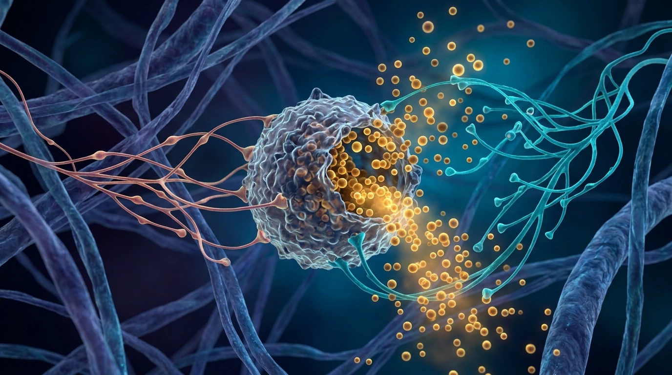
1. The Neuro-Immune Triad: MCAS, Dysautonomia, and Vagal Failure
In classical immunology, mast cells were viewed exclusively as mediators of IgE-dependent Type I hypersensitivity (anaphylaxis and common allergies). However, Mast Cell Activation Syndrome (MCAS) represents a systemic, non-IgE-mediated condition where mast cells become hypersensitive, degranulating erratically in response to environmental, emotional, and physiological stressors.
Mast cells reside in close proximity to unmyelinated autonomic nerve terminals throughout the "barrier tissues"—the skin, gastrointestinal tract, respiratory mucosa, and the dura mater of the brain. When these mast cells degranulate, they release over 200 potent inflammatory mediators, including histamine, tryptase, prostaglandins (PGD2), leukotrienes, and cytokines.
These mediators immediately interact with local autonomic nerve endings. Histamine stimulates H1, H2, and H3 receptors on perivascular sympathetic fibers and sensory afferents, driving widespread vasodilation, plasma leakage, and compensatory reflex tachycardia. For individuals navigating complex autonomic symptoms, reviewing our vagus nerve clinical foundations helps clarify this neuro-immune interplay.
2. Mast Cell Degranulation: Histamine, Tryptase & Cytokine Storms
Mast cell degranulation is not an all-or-nothing event; it can occur via "piecemeal degranulation," selectively releasing specific chemicals without cell lysis:
- Foundational Neurobiology: Review our clinical analysis on the physiological sigh and autonomic anxiety reset.
- Histamine: Binds H1 receptors causing bronchoconstriction and pruritus; H2 receptors causing gastric hyperchlorhydria and coronary vasodilation; H3 receptors in the CNS causing insomnia and microglial activation; and H4 receptors driving eosinophil chemotaxis.
- Tryptase: A neutral serine protease that cleaves extracellular matrix fibronectin, activates PAR2 receptors, and degrades connective tissues, contributing to systemic joint instability.
- Prostaglandin D2 (PGD2): Promotes profound flushing, peripheral vasodilation, headache, and bronchospasm.
- Substance P & CRH: Corticotropin-Releasing Hormone released during psychological stress binds to mast cell CRHR1 receptors, triggering direct non-allergic degranulation.
3. The Sympathetic Vicious Cycle: Adrenaline as a Mast Cell Trigger
The hallmark of dysautonomia and POTS is excessive, uncoordinated sympathetic nervous system discharge. When blood pools in the lower extremities, the brain perceives cerebral hypoperfusion and fires massive bursts of norepinephrine and epinephrine from the rostral ventrolateral medulla and adrenal glands.
Crucially, mast cells express functional beta-2 and alpha-1 adrenergic receptors on their outer membranes. While physiological low-dose adrenaline can stabilize mast cells (the basis of the EpiPen), sustained, high-intensity noradrenergic surges trigger intracellular calcium influx, provoking secondary mast cell degranulation.
This establishes an unremitting self-reinforcing vicious cycle: mast cell histamine causes vasodilation → venous return collapses → sympathetic adrenaline surges → adrenaline ruptures more mast cells → more histamine floods the circulation. Without vagal intervention, this loop traps patients in perpetual autonomic crises.
4. Biomarker & Diagnostic Profiling: MCAS vs. Typical Allergies
| Diagnostic Parameter | True Type I IgE Allergy | Mast Cell Activation Syndrome (MCAS) |
|---|---|---|
| Underlying Mechanism | IgE-mediated cross-linking on FcεRI receptors | Multifactorial non-IgE receptor activation (MRGPRX2, CRHR1, TLR) |
| Trigger Specificity | Specific allergens (peanuts, bee venom, pollen) | Multi-system triggers: foods, smells, vibration, stress, temperature |
| Serum Total / Specific IgE | Markedly elevated against specific targets | Typically normal or low |
| Serum Baseline Tryptase | Elevated in systemic mastocytosis | Often normal; episodic rise >20% + 2 ng/mL during flares |
| 24-Hour Urinary Mediators | Normal between acute events | Elevated N-methylhistamine, 11β-PGF2α, or Leukotriene E4 |
| Autonomic Involvement | Transient shock during anaphylaxis only | Chronic POTS, orthostatic intolerance, labile blood pressure, gastroparesis |
5. The Cholinergic Anti-Inflammatory Pathway as a Mast Cell Brake
The biological antidote to mast cell hyperactivity is the parasympathetic Vagus Nerve through the Cholinergic Anti-Inflammatory Pathway.
Efferent vagal fibers terminate in autonomic ganglia adjacent to mast cell niches. Acetylcholine released from cholinergic nerve terminals binds specifically to alpha-7 nicotinic acetylcholine receptors (α7nAChR) expressed on the mast cell membrane. This binding blocks intracellular calcium influx, suppresses NF-κB activation, and directly stabilizes the cell membrane, preventing granule exocytosis.
In patients with dysautonomia, resting cardiac and visceral vagal tone is profoundly depressed. Lacking adequate cholinergic tone, mast cells lose their physiological brake and enter a state of constitutive hyper-excitability. Restoring vagal nerve output is therefore a prerequisite for stabilizing refractory MCAS.
6. Comprehensive Clinical Protocol: Mast Cell & Autonomic Stabilization
Effective stabilization requires addressing histamine receptors, cellular membranes, and autonomic pacemaking concurrently:
- Receptor Blockade (H1 & H2 Antagonists): Dual receptor blockade using nonsedating H1 blockers (fexofenadine 180 mg BID or desloratadine 5 mg BID) combined with H2 blockers (famotidine 20–40 mg BID) dampens cardiovascular vasodilation and gastric hyperchlorhydria.
- Natural Mast Cell Stabilizers:
- Luteolin & Quercetin Phytosome (500 mg TID with meals): Natural flavonoids that inhibit calcium entry into mast cells and down-regulate cytokine gene expression.
- Sodium Cromoglycate (Cromolyn Sodium, 100–200 mg QID): Acts locally in the gut lumen to prevent food-triggered mucosal degranulation.
- Diaminoxidase (DAO Enzymes): Taken before meals to degrade dietary histamine in the intestinal lumen before systemic absorption.
- Sympathetic Dampening & Autonomic Reconditioning:
- Transcutaneous Auricular VNS (tVNS): 20 Hz stimulation of the left cymba conchae twice daily activates central α7nAChR pathways, directly stabilizing tissue mast cells.
- Hydration & Electrolyte Expansion: 3 to 4 liters of fluid with 5,000–7,000 mg of sodium daily expands intravascular volume, blunting the reflex sympathetic spikes that trigger mast cells.
Frequently Asked Questions (Clinical FAQ)
Why do MCAS patients frequently develop POTS and dysautonomia?
When mast cells degranulate, they flood the bloodstream with histamine and prostaglandins, which cause profound blood vessel dilation. This causes blood to pool in the abdomen and legs upon standing, forcing the heart to race (POTS) and the sympathetic system to pump out adrenaline.
Can emotional stress trigger an allergic-like MCAS flare?
Yes. Psychological stress causes the brain and nerves to release Corticotropin-Releasing Hormone (CRH) and Substance P. Mast cells express receptors for these neuropeptides and degranulate directly in response to emotional threat, completely independent of food or allergens.
What is the difference between Mastocytosis and MCAS?
Systemic Mastocytosis is a rare neoplastic disease characterized by a pathological proliferation and accumulation of abnormal mast cells (often with a KIT D816V mutation) and constantly high baseline tryptase. In MCAS, the total number of mast cells is normal, but they are hyperactive and degranulate abnormally.
How does the vagus nerve help calm histamine reactions?
The vagus nerve releases acetylcholine, which binds to alpha-7 nicotinic receptors on mast cells. This acts as a chemical brake, stabilizing the cell membrane and preventing it from rupturing and releasing histamine.
Which foods are highest in histamine and should be avoided during a flare?
Aged cheeses, fermented foods (sauerkraut, kimchi, kefir), wine, cured meats, canned fish (tuna, sardines), vinegar, tomatoes, spinach, and avocados are naturally high in histamine and should be limited during an active flare.
Why is blood tryptase often normal in patients who clearly have MCAS?
Tryptase has a very short half-life of only 2 hours and primarily spikes during severe systemic anaphylactic events. In chronic "piecemeal" MCAS, mast cells release histamine and leukotrienes without releasing massive amounts of tryptase.
Scientific References & Clinical Citations
- Mast cell activation disease: a concise practical guide for diagnostic workup and therapeutic options — Journal of Hematology & Oncology (2011). [PubMed / Study Link]
- Postural tachycardia syndrome and mast cell activation: coordinated neuro-immune autonomic dysfunction — Autonomic Neuroscience (2018). [PubMed / Study Link]
- Nicotinic acetylcholine receptor alpha 7 subunit is an essential regulator of inflammation — Nature (2003). [PubMed / Study Link]
- Histamine receptors in the nervous system: physiological and clinical implications — Frontiers in Systems Neuroscience (2016). [PubMed / Study Link]