
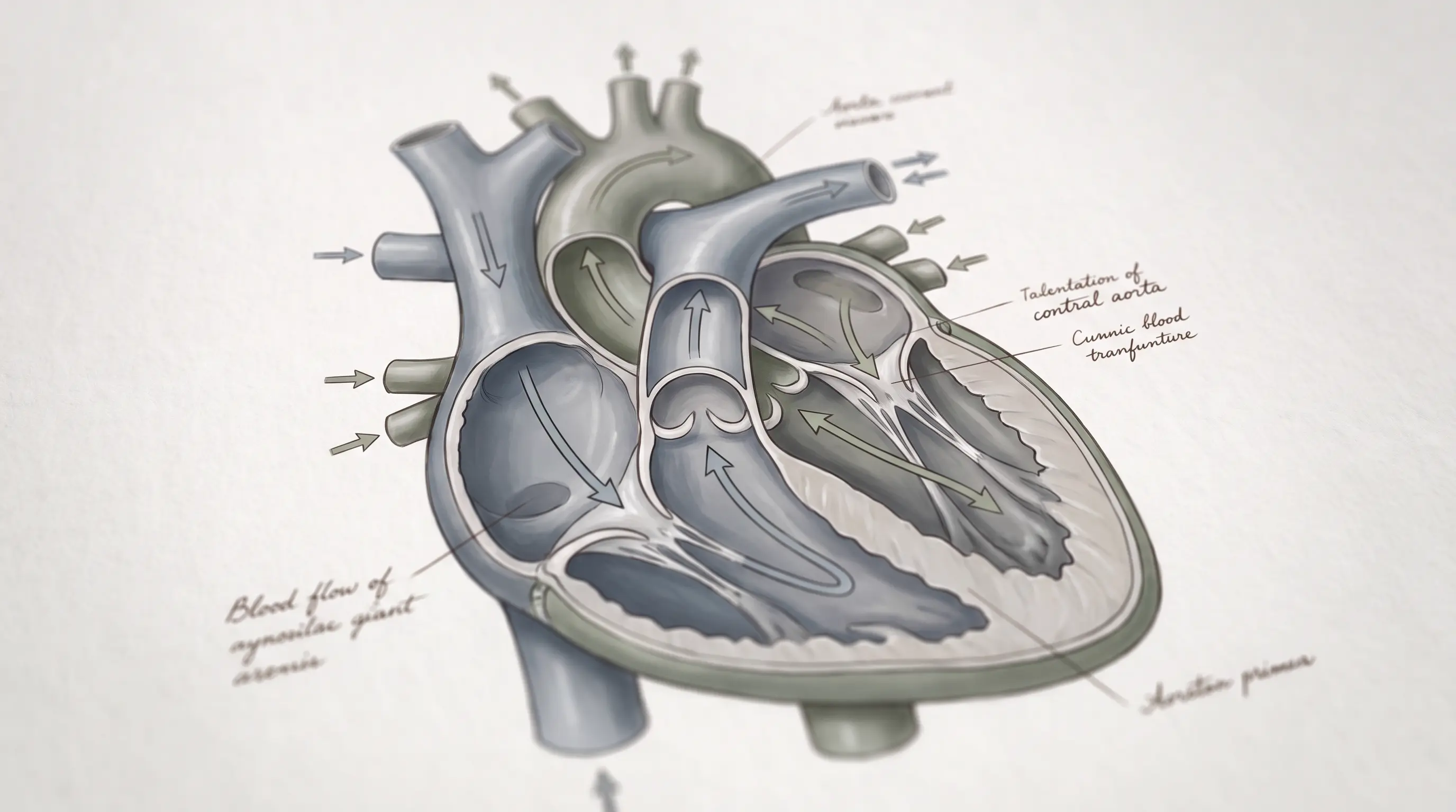
When you are told to "take a deep breath," the instruction rarely comes with an anatomy lesson. Yet the entire stress-reducing power of that breath depends on a precise chain of structures — the diaphragm, the phrenic nerve, and the vagus nerve — working in sequence to shift your nervous system out of fight-or-flight.
Diaphragmatic breathing is consistently cited as one of the most effective non-pharmacological interventions for stress, anxiety, and autonomic dysregulation (NIH/PMC, 2021). But its benefits are not psychological placebo. They are the product of measurable neuroanatomy: the way you breathe physically changes the firing rate of the vagus nerve, which in turn governs heart rate, blood pressure, digestion, and the tone of your entire parasympathetic system.
This article walks through the anatomy that makes diaphragmatic breathing work — from the dome-shaped muscle at the base of the ribcage to the cranial nerve that translates breath into calm — and explains why the specific pattern of breathing matters more than the act of breathing itself.
Key Takeaways
- The diaphragm is a dome-shaped muscle innervated by the phrenic nerve (C3-C5)
- The vagus nerve carries stretch signals from the lungs and aorta to the brainstem during each breath
- Slow breathing at 4-6 breaths per minute maximizes respiratory sinus arrhythmia and vagal outflow
- Extended exhales specifically increase parasympathetic (vagal) activation
- Heart rate variability is a direct, measurable index of vagal tone during breathing
- Consistent daily practice, not single sessions, produces durable vagal improvements
Last updated: August 3, 2026 · Reviewed by Dr. Thomas Whitaker, MD, PhD, MD
The Diaphragm: Anatomy and Function
The diaphragm is a thin, dome-shaped skeletal muscle that separates the thoracic cavity from the abdominal cavity. When it contracts, it flattens and moves downward, increasing the vertical dimension of the chest and creating negative pressure that draws air into the lungs (StatPearls/NIH, 2023).
Its structure is more complex than a simple sheet of muscle. The diaphragm consists of a central tendon surrounded by muscular fibers that originate from three distinct areas:
- Sternal portion — attaches to the posterior aspect of the xiphoid process
- Costal portion — arises from the inner surfaces of the lower six ribs and their cartilages
- Lumbar portion — originates from the lumbar vertebrae and arcuate ligaments
This three-part structure allows the diaphragm to serve two roles simultaneously: it is the primary muscle of inspiration, and it forms a crucial anatomical boundary that houses the esophagus, aorta, and — most relevant to this article — the vagus nerve, which passes through the esophageal hiatus alongside the esophagus itself.
The Phrenic Nerve vs The Vagus Nerve
Two nerves dominate the story of breathing and relaxation, and confusing them is common. They perform complementary but distinct functions:
| Feature | Phrenic Nerve | Vagus Nerve (CN X) |
|---|---|---|
| Origin | Cervical spine (C3-C5) | Brainstem (medulla) |
| Primary function | Innervates the diaphragm — drives the mechanical breath | Sensory and parasympathetic — registers breath and regulates organs |
| Direction of signal | Motor (brain to muscle) | Mostly sensory (body to brain) with motor branches to heart and gut |
| Role in relaxation | Indirect — enables the breath that triggers vagal activity | Direct — the primary parasympathetic nerve |
The phrenic nerve contracts the diaphragm, expanding the lungs. Stretch receptors in the lung tissue and aortic arch then fire along vagal afferent fibers up to the nucleus tractus solitarius (NTS) in the brainstem. The NTS responds by increasing parasympathetic output to the sinoatrial node of the heart, slowing heart rate and lowering blood pressure. In short: the phrenic moves the breath, and the vagus translates that movement into physiological calm (Frontiers in Neuroscience, 2022).
How Breathing Modulates Vagal Tone
Vagal tone refers to the continuous, tonic activity of the vagus nerve, and it is the single best physiological marker of how "relaxed" your nervous system is at any moment. Breathing modulates it through a mechanism called the Hering-Breuer reflex and through direct mechanosensory feedback.
On inhalation, the lungs inflate and stretch receptors fire, which momentarily inhibits vagal outflow to allow the heart to speed up. On exhalation, the receptors stop firing and vagal outflow increases, slowing the heart. The result is a natural oscillation — faster on the inhale, slower on the exhale — known as respiratory sinus arrhythmia.
"Slow breathing techniques are unique in producing a degree of physiological coherence — phase synchronization of heart rate, blood pressure, and respiration — that maximizes vagal afferent stimulation and central parasympathetic activation."
— Zaccaro et al., How Breath-Control Can Change Your Life: A Systematic Review, 2018
By deliberately slowing the breath, you widen the gap between the inhale and exhale oscillations, effectively amplifying the signal your vagus nerve sends to your heart and gut. This is why breathing rate matters: a stressed person breathing 18-20 times per minute produces far less vagal activation than a calm person breathing 6 times per minute.
Respiratory Sinus Arrhythmia Explained
Respiratory sinus arrhythmia (RSA) is the beat-to-beat variation in heart rate that synchronizes with the breathing cycle. It is not an arrhythmia in the pathological sense — it is a sign of healthy, flexible nervous system function.
RSA is mediated almost entirely by the vagus nerve. Higher RSA indicates stronger vagal tone and greater parasympathetic flexibility. Low or absent RSA is associated with chronic stress, depression, cardiovascular disease, and autonomic dysfunction (NIH/PMC, 2021).
The practical implication is straightforward: every heart rate variability (HRV) monitor you wear is, in essence, measuring RSA. When you practice slow diaphragmatic breathing, you are directly training the mechanism your wearable device tracks. This is why breathing exercises reliably improve HRV scores — they amplify the exact physiological signal the metric measures.
The 4-7-8 Breathing Technique
One of the most studied breathing patterns for vagal activation is the 4-7-8 technique, popularized by Dr. Andrew Weil. The extended exhale is the key: longer exhalation preferentially activates the parasympathetic nervous system.
- Inhale through the nose for a count of 4
- Hold the breath for a count of 7
- Exhale slowly and completely through the mouth for a count of 8
- Repeat for four full cycles, up to a maximum of eight
The ratio matters more than the absolute numbers. Research on breath ratios shows that exhale-to-inhale ratios of 2:1 (or longer) maximize vagal activation, while the hold phase temporarily increases sympathetic arousal before the extended exhale resets it — which is why the technique works so well for acute stress spikes (NIH/PMC, 2021).
Diaphragmatic Breathing Protocol for Vagal Health
A structured daily protocol produces the most reliable improvements in vagal tone. The following sequence takes roughly 10 minutes and can be done morning and evening:
- Position: Lie flat on your back with knees bent, or sit upright with a straight spine. Place one hand on your chest and one on your abdomen.
- Activation: Breathe so that the hand on your abdomen rises and falls while the hand on your chest stays still — this confirms diaphragmatic, not chest, breathing.
- Pacing: Target 5-6 breaths per minute. A simple pattern is inhale for 4 seconds and exhale for 6 seconds.
- Focus: Direct attention to the sensation of the breath at the navel. Lengthen the exhale progressively over the session.
- Duration: Start with 5 minutes and build to 10-20 minutes over two weeks.
Consistency outperforms intensity. Five minutes daily for eight weeks produces larger sustained improvements in heart rate variability than a single 30-minute session performed occasionally (Mayo Clinic, 2024).
How the protocol Optimizes Diaphragmatic Vagal Activation
The breathing protocol was engineered around the anatomy described above. Its core breathing missions deliberately extend the exhale phase of the breath cycle — the exact parameter that maximizes vagal outflow — and pace the breath at the 4-6 cycles-per-minute range where respiratory sinus arrhythmia peaks.
- Anatomically targeted: The audio cues coach the diaphragmatic pattern (abdominal rise, minimal chest movement) that maximizes phrenic-vagal coupling
- Extended exhale engineering: Each mission emphasizes a prolonged, controlled exhale to amplify parasympathetic drive
- Consistency scaffolding: The 28-day structure ensures the daily repetition required to raise baseline vagal tone, not just acute relaxation
The protocol does not require any special equipment, medication, or clinic visits. It is an audio-guided program that can be practiced at home in 5 to 10 minutes per session.
When to See a Doctor
You should consult a healthcare professional before starting intensive breathwork if you have:
- Chronic obstructive pulmonary disease (COPD) or asthma that is poorly controlled
- Unstable cardiovascular disease or recent heart surgery
- Recurrent fainting episodes or suspected orthostatic hypotension
- Epilepsy (some breath patterns can trigger seizures)
- Current pregnancy complications
If diaphragmatic breathing produces persistent dizziness, chest pain, or shortness of breath, stop and seek medical evaluation.