
1. Anatomy of the Diaphragm: The Musculoskeletal-Autonomic Bridge
The diaphragm is a large, dome-shaped musculotendinous sheet separating the thoracic and abdominal cavities. It anchors anteriorly to the xiphoid process, laterally to the lower six ribs and costal cartilages, and posteriorly to the lumbar vertebrae via strong muscular tendons called crura.
What makes the diaphragm physiologically unique is its dual neural wiring:
- Motor Innervation: Driven exclusively by the phrenic nerve, which originates from cervical spinal nerve roots C3, C4, and C5 ("C3, 4, 5 keeps the diaphragm alive").
- Autonomic Convergence: The central tendon and diaphragmatic crura sit in direct anatomical contact with the pericardium of the heart, the inferior vena cava, the abdominal aorta, and the vagus nerves passing through the esophageal hiatus.
Because the phrenic and intercostal nerves converge onto the same dorsal horn spinal segments as sensory fibers from the heart and gallbladder, irritation or spasms in the diaphragmatic muscle trigger identical referred pain sensations across the chest, neck, and shoulder.
2. Why Anxiety Triggers Diaphragmatic Spasms and Splinting
Under psychological threat or panic, the central nervous system activates the defensive "freeze or flight" posture. This primal somatic reflex causes dramatic biomechanical distortions in the chest:
- Inspiratory Splinting: The brain commands the body to brace against physical impact. The diaphragm contracts violently and remains locked in a semi-flattened inspiratory position, unable to relax into its resting dome configuration during exhalation.
- Accessory Muscle Overdrive: Because the diaphragm is locked in spasm, the body recruits the sternocleidomastoid, scalenes, and pectoralis minor muscles to lift the ribcage. These small neck muscles rapidly fatigue, creating burning aches radiating down into the chest.
- Alkalotic Tetany: As anxious rapid breathing blows off carbon dioxide, the resulting respiratory alkalosis drives ionized calcium levels down. This hyper-excites motor endplates, provoking spontaneous, painful tetanic twitches and sustained cramping in the diaphragmatic crura and intercostal muscles.
3. What Is True Angina? The Ischemic Cascade
To accurately distinguish functional muscle spasms from cardiovascular disease, one must understand the pathophysiology of angina pectoris. Angina occurs when myocardial oxygen demand exceeds coronary arterial oxygen supply, typically due to atherosclerotic plaque stenosis or coronary vasospasm.
Deprived of adequate oxygen, ischemic cardiomyocytes switch from aerobic metabolism to anaerobic glycolysis. Lactic acid, adenosine, and bradykinin accumulate in the cardiac interstitium, stimulating cardiac sympathetic afferent nerve endings. These signals travel through the upper thoracic sympathetic ganglia (T1-T5) into the spinothalamic tract, registering in the brain as a heavy, crushing, or burning substernal pressure.
4. Clinical Differential: Diaphragm Spasm vs. Coronary Angina
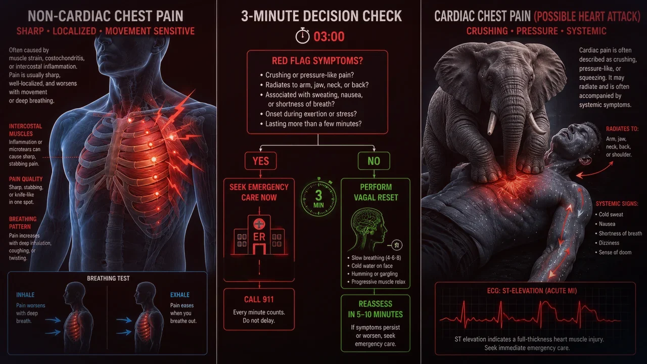
In our comprehensive clinical comparison of anxiety chest tightness vs heart attack symptoms, physicians use specific diagnostic features to separate musculoskeletal spasms from acute coronary syndrome:
| Diagnostic Parameter | Diaphragmatic / Intercostal Spasm | True Coronary Angina / Ischemia |
|---|---|---|
| Quality of Sensation | Sharp, gripping, band-like ache, stitch in side | Crushing, heavy "elephant on chest", deep vice-like squeeze |
| Effect of Respiration | Worsens dramatically on full inhale (pleuritic) | Unaffected by breathing, coughing, or inspiration |
| Effect of Posture / Movement | Changes with twisting, bending, or chest wall palpation | Completely unaffected by physical position or palpation |
| Exertional Relationship | Often occurs at rest, during anxiety, or post-stress | Triggered reliably by physical exertion; relieved by rest |
| Associated Palpitations | Accompanied by anxiety-driven autonomic palpitations | Cold sweats, severe nausea, ashen pallor, radiation to jaw |
5. Phrenic Nerve Referral: Why Diaphragm Issues Hurt the Shoulder
One of the most terrifying symptoms for anxious patients is experiencing chest tightness alongside left shoulder or neck pain—the textbook warning sign of a heart attack. However, diaphragmatic spasm causes the exact same referral pattern.
Because the phrenic nerve originates from spinal levels C3 through C5, sensory fibers from the diaphragm enter the spinal cord at the same exact level as the supraclavicular cutaneous nerves that supply sensation to the top of the shoulder and clavicle. A cramp or spasm in the left dome of the diaphragm frequently projects pain directly to the left trapezius ridge and neck, completely mimicking cardiac referral without any coronary involvement.
6. Somatic Release: Releasing Diaphragmatic Tetany Safely
When cardiac causes have been ruled out by a medical professional, patients can rapidly relieve diaphragmatic cramping using targeted neuromuscular release protocols:
- Psoas-Diaphragm Unloading Posture (Constructive Rest): Lie on your back on a firm carpet or yoga mat with knees bent at 90 degrees resting on a chair or couch. This slackens the psoas major and relieves anterior tension on the diaphragmatic crura.
- Subcostal Hooking Release: In the constructive rest posture, gently place the pads of your fingers underneath the lower edge of your ribcage. Take a slow inhale, and on the slow exhale, gently hook your fingers 1 centimeter upward under the ribs, massaging the lower costal attachments.
- Physiological Sigh Reset: Inhale deeply through the nose, immediately take a second sharp "top-off" inhale to fully stretch the alveoli and diaphragmatic fibers, then release with a long, unforced vocalized sigh through the mouth. Repeat 3 times to break tetanic motor unit firing.