
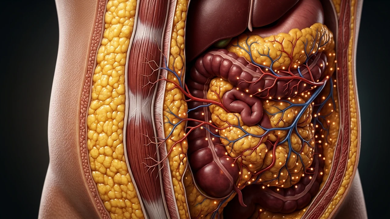
1. The Two Adipose Depots: Anatomical & Functional Divergence
Not all adipose tissue responds uniformly to caloric expenditure, exercise, or neuroendocrine signaling. Clinicians divide trunk adiposity into two fundamentally distinct anatomical compartments: subcutaneous adipose tissue (SAT), located directly beneath the dermis and fascia, and visceral adipose tissue (VAT), situated deep within the peritoneal cavity surrounding the liver, intestines, pancreas, and mesenteric vessels.
Subcutaneous fat behaves primarily as a structural, protective energy reservoir. In contrast, visceral fat operates as an aggressive, highly vascularized neuroendocrine organ. When patients experience stalled progress during fat loss protocols, they are rarely dealing with a generalized failure of lipolysis. Instead, they are experiencing selective regional lipolytic inhibition. To understand the overarching systemic barriers to abdominal weight reduction, review our core clinical analysis on why lower belly fat is so difficult to lose.
2. Receptor Kinetics: Alpha-2 vs. Beta-Adrenergic Receptors
The rate at which an adipocyte breaks down stored triacylglycerols into free fatty acids (FFAs) and glycerol is governed by the interplay between catecholamines (epinephrine and norepinephrine) and surface adrenergic receptors:
- Beta-1, Beta-2, and Beta-3 Adrenergic Receptors: These are stimulatory G-protein coupled receptors (Gs). When stimulated by catecholamines during movement or cold exposure, they activate adenylyl cyclase, elevate intracellular cyclic AMP (cAMP), and initiate protein kinase A (PKA) phosphorylation of hormone-sensitive lipase (HSL) and perilipin-1, releasing stored fatty acids into the bloodstream.
- Alpha-2 Adrenergic Receptors: These are inhibitory G-protein coupled receptors (Gi). When occupied, they inhibit adenylyl cyclase, drop intracellular cAMP levels, and aggressively halt lipolysis, preventing fat cells from emptying their contents.
In the lower abdominal subcutaneous depot and deep mesenteric visceral fat, the density of inhibitory Alpha-2 receptors is up to 300% higher than in upper body or extremity fat cells. When cortisol is chronically elevated alongside basal sympathetic tone, it upregulates Alpha-2 receptor expression while desensitizing Beta-1 and Beta-2 pathways, effectively deadlocking abdominal fat breakdown.
3. Clinical Comparison Table: Subcutaneous vs. Visceral Fat
| Biological Parameter | Subcutaneous Adipose Tissue (SAT) | Visceral Adipose Tissue (VAT) |
|---|---|---|
| Anatomical Location | Sub-dermal, superficial to muscle fascia | Omental, mesenteric, perirenal retroperitoneal |
| Glucocorticoid Receptor Density | Baseline (1x reference level) | 4x to 5x higher receptor density |
| Venous Drainage Route | Systemic circulation (inferior vena cava) | Portal vein (direct first-pass hepatic delivery) |
| 11β-HSD1 Enzymatic Activity | Low to moderate | Extremely high local cortisone-to-cortisol conversion |
| Insulin Sensitivity vs. Resistance | Moderate insulin sensitivity; buffers lipids | High basal lipolysis under insulin resistance; inflammatory |
| Adipokine & Cytokine Secretion | Predominantly adiponectin & leptin | IL-6, TNF-alpha, MCP-1, PAI-1, Angiotensinogen |
4. The 11β-HSD1 Amplification Loop in Omental Fat
The most profound molecular discriminator between stubborn belly fat and peripheral fat is the intracellular enzyme 11-beta-hydroxysteroid dehydrogenase type 1 (11β-HSD1). Circulating cortisol is largely bound to corticosteroid-binding globulin (CBG) or converted into inactive cortisone by renal 11β-HSD2.
However, visceral omental adipocytes produce enormous quantities of 11β-HSD1. This enzyme takes biologically inactive cortisone from the bloodstream and reactivates it into active cortisol inside the fat cell itself. Consequently, even if a routine serum blood draw shows "normal" total cortisol levels, the local intracellular cortisol concentration within abdominal fat can be up to 400% higher than circulating plasma levels, constantly driving adipocyte hypertrophy and visceral lipid storage.
5. Portal Circulation & The Hepatic Steatosis Cascade
Unlike subcutaneous fat, which drains directly into the systemic vena cava, visceral adipose tissue drains straight into the hepatic portal vein. When visceral adipocytes break down under abnormal stress conditions, they release a flood of toxic free fatty acids and inflammatory cytokines directly into the liver.
This portal FFA overload impairs hepatic insulin clearance, triggers hepatic gluconeogenesis (raising fasting blood glucose), and leads to metabolic dysfunction-associated steatotic liver disease (MASLD). The liver responds by packaging excess lipids into very-low-density lipoproteins (VLDL) and re-depositing them right back into the visceral omentum, creating a self-perpetuating cycle of abdominal fat accumulation.
6. The 4-Phase Protocol to Unblock Visceral Lipolysis
To reverse selective abdominal lipolysis resistance, clinical protocols must address the neuroendocrine receptor environment rather than simply cutting calories further:
- Phase 1: Blunting Nocturnal 11β-HSD1 Expression: Inhibit evening glucocorticoid signaling with targeted adaptogens (phosphatidylserine 400 mg, standardized ashwagandha withanolides) taken 60 minutes before bedtime.
- Phase 2: Restoring Deep Delta Slow-Wave Sleep: Pulsatile human growth hormone (GH), which is the body's most potent selective visceral lipolytic hormone, is secreted almost exclusively during Stages 3 and 4 of NREM sleep. Restoring autonomic parasympathetic tone at night is essential for GH release.
- Phase 3: Sub-Threshold Zone 2 Aerobic Oxidation: Keep aerobic exercise below the autonomic lactate threshold (heart rate 60–70% of max). High-intensity intervals in an already exhausted, hyper-cortisolemic individual spike ACTH and cortisol, reinforcing Alpha-2 receptor dominance.
- Phase 4: Postprandial Vagal Reactivation: Practice 5 minutes of exhalation-biased diaphragmatic breathing immediately following meals to down-regulate sympathetic vasoconstriction of splanchnic circulation, improving gut insulin sensitivity.