
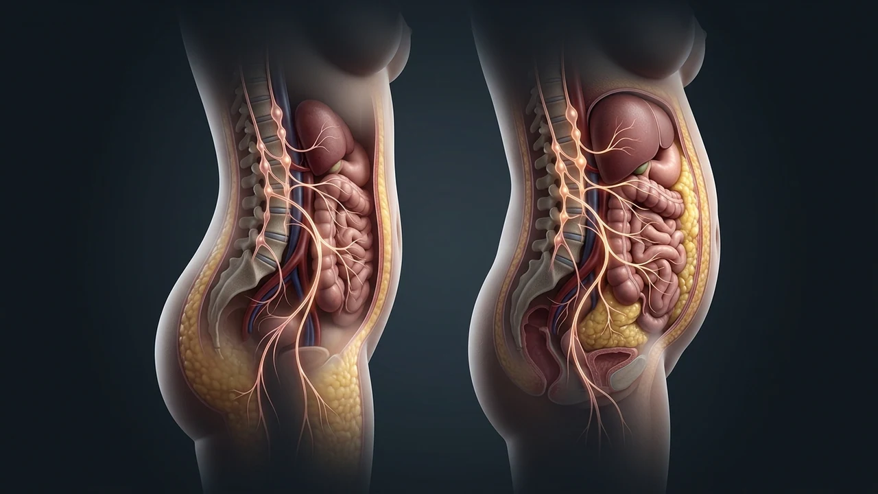
1. The Gynoid-to-Android Phenotypic Transition
Throughout female reproductive life, 17-beta estradiol (E2) orchestrates a protective, evolutionary energy storage pattern known as the gynoid distribution: fat is selectively partitioned into the gluteofemoral subcutaneous depots (hips and thighs) via high lipoprotein lipase (LPL) activity in those specific tissues.
During the perimenopausal transition, ovarian follicular depletion leads to a steep drop in circulating estradiol. Simultaneously, adipose tissue distribution transitions rapidly to an android distribution: fat deposition shifts from subcutaneous peripheral sites directly into the deep visceral omental and mesenteric compartments. The postmenopausal autonomic shift explains why stomach fat accumulates during hormonal shifts, even in women who maintain identical exercise routines and dietary habits.
2. Estrogen Receptors in Adipose Tissue: ERα vs. ERβ
Adipocytes express two distinct estrogen receptor isoforms that exert opposing influences on lipid metabolism:
- Estrogen Receptor Alpha (ERα): Expressed abundantly in healthy female adipose tissue, ERα promotes mitochondrial biogenesis, maintains insulin sensitivity, and directly suppresses Lipoprotein Lipase (LPL) in visceral fat depots. When ERα is occupied by estradiol, visceral fat accumulation is biochemically suppressed.
- Estrogen Receptor Beta (ERβ): When estradiol drops and the ERα/ERβ ratio shifts in favor of ERβ, visceral fat cells lose their protective anti-lipogenic brake, accelerating visceral adipocyte hypertrophy.
Knockout animal models lacking ERα develop severe abdominal obesity and metabolic dysfunction, demonstrating that the loss of estrogenic signaling at the adipocyte receptor level is sufficient to trigger visceral fat accumulation independent of food consumption.
3. The Autonomic Shift: Vagal Withdrawal & Vasomotor Overdrive
Estradiol is not only a reproductive hormone; it is a potent neuro-cardiovascular protectant that directly modulates central autonomic tone in the brainstem. Estradiol enhances parasympathetic vagal activity at the Nucleus Tractus Solitarius (NTS) and dampens sympathetic outflow from the rostral ventrolateral medulla (RVLM).
With the menopausal loss of estrogen:
- Resting Cardiac Vagal Tone Collapses: Heart Rate Variability (RMSSD) experiences a sharp downward inflection during perimenopause, signaling reduced vagal braking capacity.
- Sympathetic Vasomotor Tone Surges: Uninhibited central sympathetic outflow produces the classic vasomotor symptoms of hot flashes, night sweats, and resting palpitations.
- Cortisol-to-DHEA Ratio Climbs: The adrenal glands must compensate for lost ovarian steroid production, shifting steroidogenesis toward cortisol synthesis while protective androgens like DHEA-S steadily decline.
4. Clinical Comparison Table: Pre- vs. Post-Menopausal Autonomic Profile
| Physiological Parameter | Premenopausal State (E2 Dominant) | Postmenopausal State (E2 Depleted) |
|---|---|---|
| Primary Fat Storage Depot | Subcutaneous gluteofemoral (hips/thighs) | Visceral omental, mesenteric, and trunk |
| Visceral Adipocyte LPL Activity | Strongly inhibited by ERα signaling | Uninhibited; rapid triglyceride uptake |
| Heart Rate Variability (RMSSD) | Elevated; robust parasympathetic vagal buffer | Significantly reduced; sympathetic dominance |
| Nocturnal Sleep Quality | Stable Slow-Wave Sleep (SWS) architecture | Frequent awakenings via vasomotor temperature surges |
| Insulin Sensitivity in Skeletal Muscle | High; preserved GLUT4 translocation | Reduced; peripheral insulin resistance |
| Systemic Inflammatory Baseline | Low baseline C-reactive protein (CRP) | Elevated IL-6 and TNF-alpha from visceral expansion |
5. The Sleep Fragmentation & Visceral Fat Cascade
Hot flashes and night sweats are not merely uncomfortable; they are profound neuro-metabolic disruptions. A nocturnal vasomotor event is preceded by a sudden spike in sympathetic adrenaline, which drops brain temperatures and awakens the patient out of restorative Slow-Wave Sleep.
This persistent nocturnal sleep fragmentation drives daytime insulin resistance and elevates evening cortisol levels. Sleep-deprived menopausal women show marked reductions in leptin (satiety) and elevation in ghrelin (hunger), creating irresistible physiological cravings for high-glycemic carbohydrates precisely when their visceral fat cells are primed to store them.
6. Multi-Modal Clinical Strategy for Menopausal Metabolic Protection
Managing menopausal abdominal weight resistance requires addressing the neuroendocrine and autonomic environment simultaneously:
- Bioidentical Hormone Optimization: For eligible women without medical contraindications, bioidentical Hormone Replacement Therapy (transdermal 17-beta estradiol paired with micronized oral progesterone) preserves ERα signaling, halts the gynoid-to-android shift, and restores nocturnal sleep architecture.
- Targeted Vagal Neuromodulation: Transcutaneous auricular vagus nerve stimulation (taVNS) at the tragus reduces central sympathetic overdrive, dampens vasomotor frequency, and restores parasympathetic tone.
- Phytoestrogen Receptor Modulation: Incorporating dietary lignans (flaxseed) and isoflavones (genistein, daidzein) provides selective estrogen receptor modulation, partially stimulating ERα without promoting endometrial hyperplasia.
- Resistance Training for Myokine Signaling: Progressive resistance training induces skeletal muscle secretion of irisin, a myokine that promotes the "browning" of white visceral fat cells into metabolically active beige adipocytes.