
1. The Pituitary Illusion: Why Normal TSH Misses the Problem
In standard medical evaluation, thyroid function is evaluated almost exclusively through Thyroid Stimulating Hormone (TSH) secreted by the anterior pituitary gland. The pituitary monitors circulating hormones via an ultra-sensitive negative feedback loop. However, the pituitary gland possesses a fundamentally unique deiodinase enzyme profile compared to peripheral tissues like liver, muscle, and adipose tissue.
The pituitary uses Type 2 Deiodinase (D2), which is up to 1,000 times more efficient at converting T4 to active T3 than the Type 1 Deiodinase (D1) found in the rest of the body. Under high-cortisol conditions, D1 activity in peripheral fat and liver is aggressively suppressed, while pituitary D2 remains fully active. Consequently, the pituitary "senses" adequate thyroid hormone and keeps TSH within normal limits (e.g., 1.5–2.5 mIU/L), even while peripheral cells are starved of active thyroid signaling. This diagnostic trap explains why you can't lose stomach fat with normal labs.
2. Reverse T3 (rT3): The Molecular Bouncer at the Nuclear Receptor
To understand why metabolic rate collapses despite normal blood work, clinicians must analyze the pathway of Reverse T3 (rT3). In healthy individuals, T4 is converted into active Triiodothyronine (T3) by removing an iodine atom from the outer ring via 5'-deiodinase.
When cortisol is chronically elevated due to emotional stress, infection, or aggressive caloric deprivation:
- Cortisol suppresses 5'-deiodinase, starving cells of active Free T3.
- Cortisol strongly stimulates 5-deiodinase (inner ring deiodinase), shunting T4 into Reverse T3.
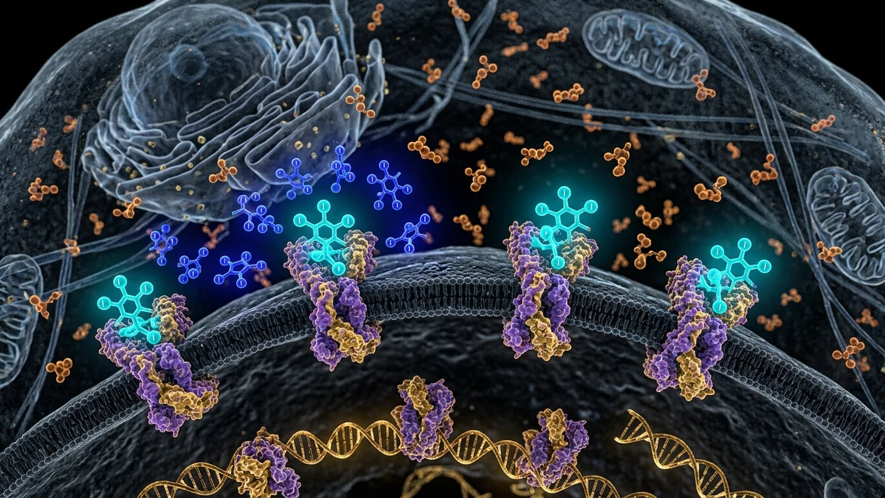
- Reverse T3 enters the cell nucleus and competitively binds to the Thyroid Hormone Receptor (THR). Because rT3 has zero metabolic activity, it functions as a competitive antagonist—blocking real T3 from initiating gene transcription for mitochondrial uncoupling and lipid oxidation.
3. Clinical Comparison: Serum Blood Tests vs. Cellular Thyroid Reality
| Diagnostic Marker | Standard Laboratory Result | Actual Cellular / Physiological Reality Under High Cortisol |
|---|---|---|
| TSH (Thyroid Stimulating Hormone) | 1.8 mIU/L ("Normal") | Pituitary D2 enzyme maintains local feedback; blind to peripheral deficit. |
| Free T4 (Thyroxine) | 1.2 ng/dL ("Normal") | Adequate pro-hormone in blood, but cannot be activated in target cells. |
| Free T3 (Triiodothyronine) | Low-normal or suboptimal | Markedly reduced intracellular T3/rT3 ratio in liver and adipose tissue. |
| Reverse T3 (rT3) | Rarely tested by standard panels | Elevated (> 20 ng/dL); actively occupying and blocking nuclear receptors. |
| Basal Oral Temperature | Unmeasured in clinic | Consistently low (96.8°F – 97.4°F), proving depressed metabolic heat generation. |
| Adipose Lipolysis Rate | Unmeasured in clinic | Severely blunted in visceral omentum; high triglyceride accumulation. |
4. Cortisol's Direct Suppression of Thyroid Receptor Expression
Even if Free T3 levels manage to remain adequate in the bloodstream, elevated glucocorticoids exert a direct inhibitory effect on the transcription of the thyroid hormone receptor itself. Research in neuroendocrinology reveals that cortisol down-regulates Thyroid Hormone Receptor Alpha-1 (TRα1) and Beta-1 (TRβ1) mRNA expression in visceral adipocytes and skeletal muscle.
This creates a state of cellular thyroid resistance: the hormone is circulating in the blood, but the receptors on cell nuclei are diminished in number and unresponsive. The patient exhibits classic hypothyroid symptoms—stubborn abdominal weight gain, dry skin, thinning outer eyebrows, constipation, and brain fog—while their doctor insists "your thyroid is completely healthy."
5. The Free T3 to Reverse T3 Ratio: The Golden Clinical Marker
To accurately assess cellular thyroid metabolism, clinicians should calculate the Free T3 to Reverse T3 ratio (using identical measurement units):
A healthy, metabolically active cellular ratio of Free T3 (pg/mL) ÷ Reverse T3 (ng/dL) × 100 should exceed 20. When this ratio falls below 15, or especially below 10, the patient is in a state of pronounced functional intracellular hypothyroidism driven by glucocorticoid excess, regardless of what their TSH reads.
6. The 4-Step Clinical Protocol to Clear Reverse T3 & Restore Metabolism
Clearing reverse T3 and restoring thyroid sensitivity requires turning down the adrenal alarm while supporting peripheral deiodinase enzymes:
- Eliminate Endocrine Starvation: Ensure adequate daily carbohydrate consumption (minimum 120–150g for active women, 150–200g for men). Hepatic deiodinase enzymes require glycogen to function; chronic low-carb diets in stressed individuals are the #1 driver of high rT3.
- Deiodinase Trace Mineral Co-Factors: Supplement with 200 mcg of L-selenomethionine and 30 mg of zinc glycinate. Selenium is an absolute structural component of the deiodinase enzyme molecule; without it, T4 conversion to rT3 is favored.
- HPA Axis Quenching: Use 400 mg of standardized phosphatidylserine and 300 mg of KSM-66 ashwagandha in the late afternoon to blunt excessive ACTH signaling to the adrenal cortex.
- Vagal Parasympathetic Tone Restoration: Practice heart rate variability biofeedback for 10 minutes daily at the 0.1 Hz resonance frequency (6 breaths per minute) to lower sympathetic vasoconstriction of the thyroid and liver.