
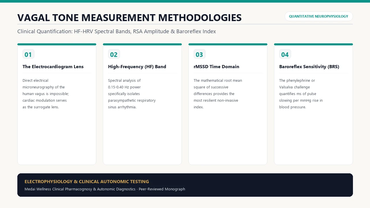
1. Defining Vagal Tone: Why Direct Measurement Is Difficult
In modern functional medicine and neurology, "increasing vagal tone" has become the preeminent goal for patients recovering from chronic anxiety, dysautonomia, and trauma. But how do neurophysiologists actually measure vagal tone with scientific precision?
Unlike skeletal motor nerves, where an electromyography (EMG) needle can record direct electrical action potentials, the human vagus nerve sits deep inside the carotid sheath of the neck, running directly beside the carotid artery and internal jugular vein. Inserting a microneurography needle into the cervical vagus carries catastrophic risks of carotid rupture or vagal cardiac standstill.
Consequently, autonomic neuroscientists measure the biological footprint of the vagus nerve on its primary target organ: the Sinoatrial (SA) node of the human heart.
2. Spectral Frequency Domain: The 0.15–0.40 Hz High-Frequency Band
When continuous electrocardiographic (ECG) data is subjected to Fast Fourier Transform (FFT) or autoregressive spectral analysis, total heart rate variability is divided into distinct frequency bands:
- Very-Low-Frequency (VLF: 0.0033–0.04 Hz): Reflects long-term thermoregulation, renin-angiotensin-aldosterone fluctuations, and hepatic metabolism.
- Low-Frequency (LF: 0.04–0.15 Hz): Reflects a mixture of sympathetic and parasympathetic influence, heavily modulated by the 10-second Mayer waves of arterial baroreceptors.
- High-Frequency (HF: 0.15–0.40 Hz): Pure parasympathetic vagal modulation. Because the biochemical clearing of norepinephrine requires several seconds, sympathetic signaling is too sluggish to operate above 0.15 Hz. Acetylcholine released by the vagus nerve acts and hydrolyzes within 50 to 100 milliseconds, allowing it to oscillate at respiratory frequencies (9 to 24 breaths per minute). Thus, HF power is 100% abolished by surgical vagotomy or intravenous atropine.
3. Respiratory Sinus Arrhythmia (RSA): The Porges-Bohrer Method
Developed by Dr. Stephen Porges (originator of Polyvagal Theory) and Dr. Craig Bohrer, the Porges-Bohrer algorithm extracts the exact amplitude of Respiratory Sinus Arrhythmia (RSA) while mathematically filtering out slow baseline drifts and respiratory rate distortions:
In this method, the patient breathes at a controlled pace while continuous ECG records the precise millisecond delta between the peak heart rate during inhalation and the trough heart rate during exhalation. A larger peak-to-trough gap indicates robust, resilient cardioinhibitory vagal tone.
4. Baroreflex Sensitivity (BRS): The Cardiovascular Stress Test
In specialized hospital autonomic laboratories (such as the Mayo Clinic or Cleveland Clinic), doctors test dynamic vagal tone using Baroreflex Sensitivity (BRS) testing:
- The patient performs a Valsalva maneuver: blowing into a mouthpiece connected to a pressure gauge at 40 mmHg for 15 seconds, followed by sudden release.
- As arterial blood pressure spikes upon release (Phase IV overshoot), stretch receptors in the carotid sinus fire impulses into the solitary tract.
- The laboratory computers calculate the BRS slope: how many milliseconds of cardiac deceleration (pulse slowing) occur for every 1 mmHg rise in systolic blood pressure (\( ext{ms/mmHg}\)).
- A normal BRS slope is \(> 10 ext{ ms/mmHg}\). A depressed slope (< \(3 ext{ ms/mmHg}\)) signifies severe vagal baroreflex failure and heightened cardiovascular mortality risk.
5. Clinical Comparison Table of Vagal Measurement Tools
| Measurement Modality | Primary Metric | Clinical Accuracy | Equipment Required |
|---|---|---|---|
| High-Frequency Spectral Analysis (HF) | HF Power (\( ext{ms}^2\)) in 0.15–0.40 Hz band | Gold Standard for resting tone | Medical ECG with specialized frequency software |
| Porges-Bohrer RSA Algorithm | RSA Amplitude (\(\ln( ext{ms}^2)\)) | Gold Standard for developmental/psychology trials | Synchronized ECG + respiratory chest belt |
| Time-Domain rMSSD | rMSSD (milliseconds) | Excellent surrogate for HF power (r > 0.90) | Chest strap monitor (Polar H10) or validated ring |
| Baroreflex Sensitivity (BRS) | \( ext{ms/mmHg}\) slope during Valsalva | Gold Standard for dynamic baroreflex reserve | Continuous beat-to-beat Finapres blood pressure monitor |
6. How to Measure Your Vagal Tone at Home Accurately
You can achieve research-grade autonomic tracking at home without an expensive hospital visit:
- Hardware: Acquire a clinical-grade Bluetooth heart rate chest strap (such as the Polar H10), which records true electrical R-peaks with millisecond accuracy (optical wrist sensors are prone to motion artifact).
- Software: Use validated HRV analysis apps like Elite HRV or Kubios HRV.
- The Morning Readiness Protocol: Upon waking, empty your bladder, sit quietly in a comfortable chair for 2 minutes, and then record a 5-minute stationary measurement while breathing naturally. Note your rMSSD and natural log of rMSSD (Ln rMSSD).
Frequently Asked Questions
Why is rMSSD considered a reliable proxy for High-Frequency (HF) HRV?
Mathematical correlation studies prove that rMSSD and HF spectral power correlate at greater than \(r = 0.90\). Because successive beat differences are calculated within milliseconds, they inherently isolate the fast-acting acetylcholine signaling of the vagus nerve.
Does a slow resting heart rate always mean high vagal tone?
Not necessarily. While high vagal tone naturally slows the resting heart rate via SA node hyperpolarization, an abnormally slow heart rate (bradycardia < 50 bpm) can also stem from intrinsic conduction disease, hypothyroidism, or electrolyte imbalances.
Can vagal tone be improved in adults?
Yes. Autonomic neuroplasticity is lifelong. Structured interventions—including daily resonance frequency breathing, zone 2 aerobic conditioning, transcutaneous auricular VNS (aVNS), and cold face immersion—consistently produce measurable increases in resting rMSSD and HF power within 6 to 12 weeks.