
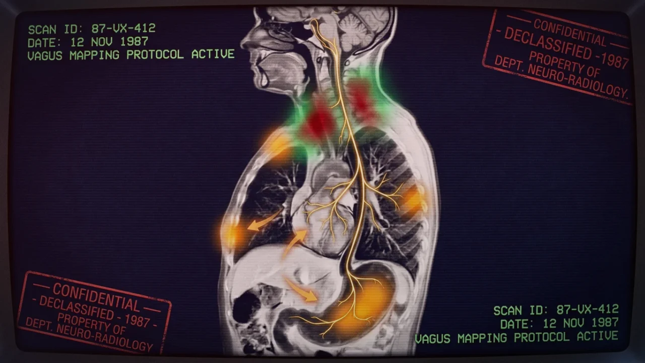
1. What Are Esophageal Spasms? Peristalsis Gone Haywire
The human esophagus is a 25-centimeter muscular conduit divided into two distinct anatomical zones: the upper one-third consists of striated skeletal muscle innervated directly by somatic motor vagal fibers from the nucleus ambiguus, while the lower two-thirds consists of involuntary smooth muscle governed by the myenteric plexus (Auerbach’s plexus) and the autonomic nervous system.
In healthy primary peristalsis, swallowing initiates a coordinated, sequential wave of contraction: circular muscles squeeze behind the food bolus while longitudinal muscles shorten, driven by acetylcholine, while the muscle immediately ahead of the bolus and the lower esophageal sphincter (LES) completely relax. In distal esophageal spasm (DES), this sequential choreography disintegrates: the smooth muscle of the distal esophagus contracts simultaneously, tortuously kinking the organ into what radiographically appears as a “corkscrew esophagus.”
2. High-Resolution Manometry: Defining Jackhammer and Nutcracker Patterns
The diagnosis of esophageal motility disorders was revolutionized by High-Resolution Manometry (HRM) utilizing the Chicago Classification v4.0. A specialized transnasal catheter containing 36 circumferential pressure sensors measures pressure topographies during water swallows:
- Distal Esophageal Spasm (DES): Defined by premature contractions with a distal latency of less than 4.5 seconds in at least 20% of swallows. The contraction wave travels too fast, jamming the esophagus before food can pass.
- Hypercontractile (Jackhammer / Nutcracker) Esophagus: Characterized by massive, repetitive hyper-contractile waves. While normal peristalsis generates contraction pressures of 30 to 100 mmHg, Jackhammer esophagus produces pressures exceeding 200 to 300 mmHg, with a Distal Contractile Integral (DCI) exceeding 8,000 mmHg·s·cm. These violent pressures compress intramural nerve fibers and micro-vessels, provoking excruciating ischemic substernal pain.
3. The Nitric Oxide Collapse: How Stress Provokes Spasms
Why do esophageal spasms flare up violently during episodes of acute anxiety, emotional confrontations, or panic attacks? Normal orderly peristalsis depends on a delicate temporal balance between postganglionic excitatory cholinergic neurons (releasing acetylcholine) and inhibitory NANC neurons (releasing nitric oxide and VIP).
Nitric oxide (NO) is the essential physiological "clutch" that delays contraction and relaxes the smooth muscle. Under acute sympathetic hyperarousal, splanchnic adrenergic surges and oxidative stress degrade neuronal nitric oxide synthase (nNOS). Lacking the inhibitory calming brake of nitric oxide, cholinergic excitatory impulses run completely unchecked. When the patient swallows—or even swallows saliva while hyperventilating—the circular smooth muscle layer locks into a sustained, tetanic cramp.
4. The Cardiac Mimic: Shared Sensory Wiring with Myocardial Angina
The primary reason esophageal spasms induce absolute panic is that they stimulate the exact same neurological alarm circuits as an ischemic heart attack. In our comprehensive medical analysis of non-cardiac esophageal chest pain, we detail the neuroanatomy of visceral sensory convergence:
Both cardiac sensory afferents and esophageal sensory afferents travel through the sympathetic cardiac and splanchnic nerves to synapse onto second-order neurons in the T1 through T5 dorsal root ganglia and dorsal horn of the spinal cord. When high-pressure esophageal spasms stimulate mechanoreceptors, the spinal cord cannot differentiate whether the nociceptive signal originated in the left anterior descending coronary artery or the distal esophagus. The pain is projected somatotopically to the substernal chest, the left arm, the jaw, and the interscapular back.
The Nitroglycerin Confounder
In emergency triage, patients often report that taking a sublingual nitroglycerin tablet or a calcium channel blocker instantly relieved their crushing chest pain, reinforcing their conviction that they suffered an anginal heart attack. However, because both the coronary arteries and the lower esophagus are composed of smooth muscle, nitric oxide donors and calcium channel blockers relax esophageal spasms just as effectively as coronary vasospasms. Response to nitroglycerin does NOT prove a cardiac origin!
5. Distinguishing Esophageal Spasms from Acute Coronary Syndromes
While every acute episode of severe chest pain demands immediate emergency cardiac rule-out (ECG and troponin), several clinical nuances distinguish esophageal spasms once safety is established:
| Clinical Feature | Esophageal Spasm / Nutcracker | True Coronary Ischemia / Angina |
|---|---|---|
| Trigger Factor | Swallowing hot/cold liquids, emotional distress, eating | Physical exertion (climbing stairs, running), walking uphill |
| Associated Dysphagia | Difficulty swallowing, sensation of food stuck in chest | Swallowing completely normal and unobstructed |
| Duration of Episode | Variable (spasms can pulse for seconds or hours) | Stable angina: 2–10 min; Infarction: persistent > 20 min |
| Electrocardiogram (ECG) | Completely normal during active pain | ST-segment elevation/depression, T-wave inversion |
6. Neuromuscular Reset: Alleviating Hypercontractile Spasms
Managing esophageal motility disorders requires reducing esophageal smooth muscle tone and restoring vagal inhibitory neurotransmission:
- Warm Water Swallows: Ingesting small sips of warm water (around 40°C / 104°F) during an acute spasm relaxes the smooth muscle and stimulates primary peristaltic clearance, whereas cold ice water triggers severe spasms in sensitive individuals.
- Peppermint Oil (Menthol): Peppermint oil acts as a natural L-type calcium channel blocker on esophageal smooth muscle cells. Dissolving 2 to 3 drops of pure culinary peppermint oil in 2 ounces of warm water immediately before meals has been shown in clinical manometry studies to eliminate simultaneous spastic contractions.
- Low-Dose Smooth Muscle Relaxants: Sildenafil (phosphodiesterase-5 inhibitor) preserves intracellular cGMP, mimicking nitric oxide and relaxing esophageal muscle during severe refractory spasms.
- Autonomic Diaphragmatic Anchoring: Performing slow, unforced nasal breathing with soft abdominal expansion relieves phrenic nerve tension around the esophageal hiatus, preventing mechanical impingement on the lower esophagus.