
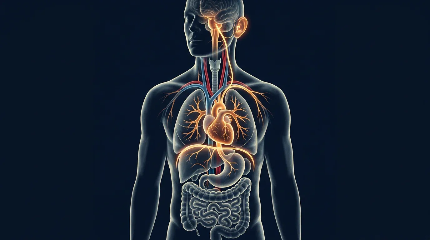
From lowering your heart rate after a sudden scare to directing gut motility and dampening systemic inflammation, the vagus nerve is the biological foundation of resilience, stress recovery, and emotional regulation. Understanding its anatomy, function, and clinical significance provides the master key to nervous system health.
Anatomy & Pathway: Where Does the Vagus Nerve Travel?
The vagus nerve is actually a paired structure, consisting of a left and right branch originating in the medulla oblongata of the brainstem. It exits the cranium through the jugular foramen and travels down the carotid sheath alongside the internal jugular vein and carotid artery.
As detailed in neurological literature from StatPearls / National Library of Medicine, the vagus nerve branches to innervate vital structures across four distinct anatomical zones:
| Anatomical Region | Vagal Nerve Branches | Primary Biological Functions | Clinical Relevance |
|---|---|---|---|
| Cervical (Neck) | Superior & recurrent laryngeal nerves, pharyngeal branches, cardiac branches | Controls swallowing, vocal cord movement, throat sensations, heart rate (SA/AV nodes) | Voice changes, dysphagia, arrhythmias, carotid sinus hypersensitivity |
| Thoracic (Chest) | Cardiac & pulmonary plexuses, esophageal branches | Slows heart rate (vagal brake), controls bronchial constriction, esophageal peristalsis | Bronchospasm, esophageal dysmotility, vasovagal syncope |
| Abdominal (Gut) | Celiac, hepatic, gastric, pancreatic, splenic, intestinal branches (subdiaphragmatic plexus) | Regulates stomach acid, digestive peristalsis, satiety signals, bile release, blood glucose | Gastroparesis, functional dyspepsia, IBS, bile reflux, glycemic variability |
| Auricular (Ear) | Arnold's nerve (auricular branch) | Provides sensory innervation to the external ear canal, tragus, and concha | Transcutaneous VNS (tVNS) access point; cough reflex from ear cleaning |
Left vs. Right Vagus Nerve: Functional Asymmetry
The left and right vagus nerves follow slightly different paths and have distinct organ emphasis:
- Right Vagus: Primarily innervates the sinoatrial (SA) node—the heart's primary pacemaker. Right vagal stimulation predominantly affects heart rate. Travels posterior to the esophagus.
- Left Vagus: Primarily innervates the atrioventricular (AV) node—controlling conduction velocity. Left vagal stimulation affects heart rhythm and AV block risk. Travels anterior to the esophagus.
- Gut Innervation: Both contribute to the esophageal plexus, but the left vagus forms the anterior gastric plexus and the right forms the posterior gastric plexus. The right vagus gives off the celiac branch supplying the liver, pancreas, and proximal small intestine.
80/20 Bidirectional Signaling: Body-to-Brain Communication
A widespread misconception is that the vagus nerve primarily sends commands from the brain down to the body. In reality, 80% of vagal nerve fibers are afferent (sensory), sending continuous data from gut visceral receptors, heart baroreceptors, and immune signals upward to the brainstem.
Only 20% of fibers are efferent (motor), carrying brain commands down to regulate heart rhythm, secretion, and digestion. This means that altering your physical state—such as through diaphragmatic breathing or cold exposure—directly changes your brain's perception of threat and safety.
Afferent Pathways: The Information Superhighway
Vagal afferents terminate primarily in the nucleus of the tractus solitarius (NTS) in the medulla, which then projects to:
- Parabrachial nucleus → amygdala, hypothalamus (emotional/autonomic integration)
- Thalamus → insular cortex (interoceptive awareness)
- Locus coeruleus → norepinephrine modulation (arousal)
- Dorsal raphe → serotonin modulation (mood)
- Ventral tegmental area → dopamine modulation (reward/motivation)
This explains why gut inflammation, heart arrhythmias, or liver dysfunction can directly cause anxiety, depression, and cognitive fog—the brain is literally "reading" the body's distress signals via the vagus nerve.
The Cholinergic Anti-Inflammatory Pathway
One of the most revolutionary scientific discoveries in neuro-immunology is the vagus nerve's role in controlling systemic inflammation. When vagal efferent fibers release the neurotransmitter acetylcholine, it binds to alpha-7 nicotinic acetylcholine receptors (α7nAChR) on macrophages in the spleen and gut.
This binding inhibits the release of pro-inflammatory cytokines such as TNF-alpha, Interleukin-1β (IL-1β), Interleukin-6 (IL-6), and High Mobility Group Box 1 (HMGB1). Consequently, high vagal tone acts as a natural, non-pharmaceutical anti-inflammatory shield. Learn more in our article on vagus nerve and immune inflammation.
Clinical Implications of the CAP
| Condition | Vagal Tone Status | Inflammatory Markers | VNS Therapeutic Potential |
|---|---|---|---|
| Rheumatoid Arthritis | Low | High TNF-α, IL-6 | Implantable VNS shows symptom reduction |
| Inflammatory Bowel Disease | Low | High TNF-α, IL-1β, HMGB1 | tVNS + medication enhances remission |
| Sepsis/Septic Shock | Acute failure | Cytokine storm | Emerging: VNS as adjunct to reduce mortality |
| Post-COVID Syndrome | Low (dysautonomia) | Persistently elevated IL-6, CRP | tVNS trials ongoing for fatigue/brain fog |
| Major Depressive Disorder | Low | Elevated CRP, IL-6 | FDA-approved implantable VNS for TRD |
10 Essential Facts About the Vagus Nerve
- It acts as the heart's natural brake: Without vagal inhibition, the heart's intrinsic pacemaker (SA node) would fire at an unsustainable 100+ beats per minute at rest. The vagus nerve provides continuous "braking" tone, allowing heart rate to settle at 60–80 bpm. This vagal brake can be rapidly released (sympathetic surge) or engaged (parasympathetic recovery).
- It directly regulates Heart Rate Variability (HRV): High HRV reflects robust, flexible vagal tone and excellent stress recovery capacity. HRV is the gold-standard non-invasive biomarker for vagal function. Learn how to track HRV in our guide to heart rate variability and vagal tone.
- It powers the gut-brain axis: Vagal fibers coordinate gastric motility, enzyme secretion, and nutrient sensing. Weak vagal tone can cause gastroparesis, functional dyspepsia, and small intestinal bacterial overgrowth (SIBO) via impaired migrating motor complex (MMC).
- It can be stimulated through the ear: The auricular branch (Arnold's nerve) allows non-invasive transcutaneous vagus nerve stimulation (tVNS) through the ear tragus and concha. This is the basis for consumer tVNS devices and clinical neuromodulation.
- It influences vocal tone and speech: The recurrent laryngeal nerves (vagal branches) modulate pitch, volume, and voice resonance during social interaction. Voice changes (hoarseness, breathiness, monotone) can signal vagal dysfunction.
- It mediates the diving reflex: Cold water applied to the face (especially forehead, cheeks, nose) instantly activates the trigeminal-vagal reflex, dropping heart rate by 15–25% within seconds. This is the fastest known non-drug vagal activation method.
- It controls the globus sensation: Pharyngeal and cricopharyngeal muscle spasms caused by autonomic imbalance create the feeling of a lump in the throat—often misdiagnosed as structural pathology.
- It is central to Polyvagal Theory: Dr. Stephen Porges demonstrated that the vagus nerve has two distinct branches—myelinated ventral vagal (social engagement, safety) and unmyelinated dorsal vagal (immobilization, freeze). This phylogenetic distinction revolutionized trauma treatment.
- It can be strengthened like a muscle: Regular somatic practices (breathing, cold exposure, humming, gargling, neck mobility) increase baseline vagal tone over weeks to months. This is autonomic conditioning—analogous to cardiovascular fitness but for the parasympathetic system.
- It works synergistically with neurotransmitters: Vagal activation increases GABA release in the amygdala (anxiety reduction), modulates serotonin in the raphe nuclei (mood stabilization), and influences dopamine in the reward pathway (motivation). This makes the vagus nerve a master regulator of the neurochemical milieu.
Measuring Vagal Tone: Clinical & Consumer Tools
| Method | Invasiveness | Accuracy | Cost | Clinical Use |
|---|---|---|---|---|
| ECG-based HRV (RMSSD, HF-HRV) | Non-invasive | Gold Standard | Low (wearables) | Autonomic assessment, treatment tracking |
| Heart Rate Recovery (HRR) post-exercise | Non-invasive | High | Free | Cardiac vagal reactivation capacity |
| Baroreflex Sensitivity (BRS) | Minimally invasive | Very High | High (lab) | Research, cardiac risk stratification |
| Pupillometry (Pupil Light Reflex) | Non-invasive | Moderate-High | Moderate | Vagal vs. sympathetic balance |
| Exhaled Breath CO2 / Respiratory Sinus Arrhythmia | Non-invasive | Moderate | Low | Breathing-vagal coupling assessment |
Symptoms of Vagus Nerve Dysfunction
When the vagus nerve becomes impaired due to chronic stress, viral infections (EBV, COVID-19), physical trauma (whiplash, surgery), or metabolic disease (diabetes), symptoms manifest across multiple body systems. Common indicators include:
- Cardiovascular: Inappropriate sinus tachycardia, orthostatic intolerance, vasovagal syncope, arrhythmias
- Digestive: Gastroparesis, bloating, nausea, constipation, SIBO, difficulty swallowing
- Neurological: Brain fog, fatigue, exercise intolerance, temperature dysregulation
- Psychiatric: Anxiety, depression, PTSD exacerbation, panic attacks
- Immune: Frequent infections, slow wound healing, autoimmune flares, chronic inflammation
- Voice/Throat: Hoarseness, globus sensation, chronic cough, choking sensation
Read our comprehensive analysis of vagus nerve dysfunction symptoms.
Vagus Nerve & Long COVID / Post-Viral Syndromes [VERIFICAR FONTE]
Emerging research suggests that SARS-CoV-2 may directly infect vagal afferents via ACE2 receptors in the nodose ganglion, or trigger autoimmune cross-reactivity against vagal tissue. This could explain the high prevalence of dysautonomia (POTS, orthostatic intolerance), gastroparesis, and chronic fatigue in Long COVID. tVNS and autonomic rehabilitation protocols are currently in clinical trials for post-viral vagal neuropathy.
Medical Disclaimer: This article is for informational purposes only and does not substitute professional medical advice or diagnosis. Consult a qualified physician for neurological or autonomic health concerns. Vagus nerve stimulation devices should be used under medical supervision.