
The Biophysics of Frequency: Matching Neural Firing Rates
Axons within peripheral nerves do not respond identically to all electrical frequencies. The Auricular Branch of the Vagus Nerve (ABVN) consists predominantly of medium-to-large diameter, myelinated A-beta (Aβ) sensory fibers, with a smaller population of unmyelinated C-fibers. A-beta fibers possess refractory periods between 1.0 and 2.5 milliseconds, allowing them to follow electrical stimulation frequencies up to roughly 50Hz without conduction block or synaptic fatigue.
When an external electrical pulse depresses the membrane potential of an Aβ axon below its threshold, an action potential propagates orthodromically toward the brainstem. The frequency of these incoming spikes dictates how downstream second-order neurons in the Nucleus Tractus Solitarius (NTS) integrate and route the signal:
- Low Frequency (0.5 - 5 Hz): Intermittent, low-rate sensory signaling that often mimics baseline visceral interoceptive tone. Highly tolerable, but generally insufficient to recruit neuroplastic changes in cortical networks.
- Medium Frequency (20 - 30 Hz): The physiological "sweet spot." Matches the natural burst firing rates of medullary catecholaminergic neurons, eliciting sustained neurotransmitter release without receptor desensitization.
- High Frequency (50 - 100+ Hz): Tends to induce rapid synaptic fatigue at the first laryngeal/auricular relay synapse, frequently causing cutaneous pain or local muscle spasm without systemic benefit.
20Hz: The Central Neuro-Psychiatric Target (Locus Coeruleus & Amygdala)
The landmark trials validating taVNS for major depressive disorder, post-traumatic hypervigilance, and cognitive rehabilitation converge on 20Hz. The neurobiological mechanism centers on the ascending projections from the NTS to the Locus Coeruleus (LC) in the rostral pons.
The Locus Coeruleus is the principal site of norepinephrine (NE) synthesis in the central nervous system. When stimulated at 20Hz, NTS excitatory glutamatergic projections provoke the LC to fire in a sustained, tonic-phasic hybrid pattern. This elevates extracellular norepinephrine and dopamine in the prefrontal cortex and hippocampus, which:
- Promotes Long-Term Potentiation (LTP) and Brain-Derived Neurotrophic Factor (BDNF) expression, enhancing neuroplasticity.
- Exerts inhibitory GABAergic control over the central nucleus of the amygdala, diminishing panic responses and fearful memory consolidation.
- Modulates the default mode network (DMN), interrupting repetitive negative thinking and ruminative depressive loops.
25Hz - 30Hz: The Peripheral Cardiovascular & Anti-Inflammatory Target
When the clinical objective shifts from neuropsychiatric modulation to autonomic cardiovascular recalibration—such as increasing high-frequency HRV, augmenting baroreflex sensitivity, or activating the Cholinergic Anti-Inflammatory Pathway—frequencies between 25Hz and 30Hz demonstrate clear superiority.
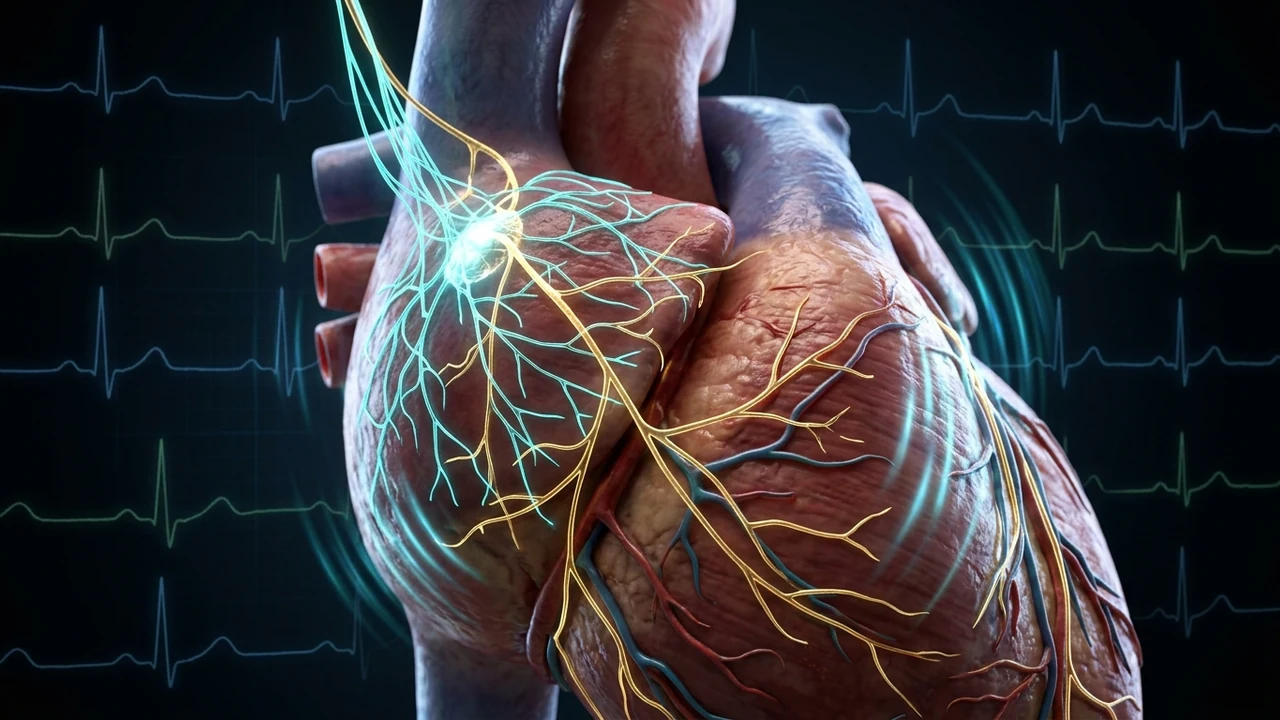
The reason lies in the anatomical wiring of the Nucleus Ambiguus. Motor neurons within the nucleus ambiguus, which project efferent myelinated B-fibers directly to the cardiac ganglia, naturally fire in bursts synchronized with the cardiac and respiratory cycles. Laboratory testing demonstrates that delivering 25Hz to 30Hz bursts to auricular afferents produces the highest rate of synaptic integration within the nucleus ambiguus, prompting maximum acetylcholine release at the cardiac sinoatrial node. This produces a measurable elevation in Root Mean Square of Successive Differences (RMSSD) and strengthens the baroreflex arc, as documented in our detailed clinical monograph on baroreflex sensitivity and autonomic blood pressure stabilization.
| Clinical Parameter | 20 Hz Protocol | 25 - 30 Hz Protocol |
|---|---|---|
| Primary Target Nuclei | Locus Coeruleus, Dorsal Raphe, Amygdala | Nucleus Ambiguus, Dorsal Motor Vagal Nucleus |
| Neurochemical Output | Central Norepinephrine, Serotonin, BDNF | Peripheral Acetylcholine (ACh), Nitric Oxide |
| Primary Indications | Depression, Hyperarousal, Brain Fog, PTSD | Hypertension, Low HRV, Dysautonomia, Gut Inflammation |
| Standard Duty Cycle | 30 seconds ON / 30 seconds OFF | Continuous or 60s ON / 30s OFF |
| Recommended Pulse Width | 250 μs (rectangular biphasic) | 200 - 300 μs (rectangular biphasic) |
Pulse Width Considerations: Why 200-300 μs Protects Patient Comfort
Frequency dictates how many times per second a pulse is delivered, but pulse width determines how long each individual pulse lasts. If the pulse width is too short (<50 μs), high voltage is required to deposit sufficient charge, creating cutaneous stinging. Conversely, if the pulse width exceeds 500 μs, unmyelinated C-fibers (pain conductors) and motor axons are activated, causing involuntary muscular contractions and pain.
Research by Badran and colleagues at the Medical University of South Carolina (MUSC) confirmed that a pulse width of 200 to 300 microseconds (μs) provides optimal charge injection into A-beta fibers while keeping current below the sensory pain threshold. Clinicians should ensure their devices generate charge-balanced biphasic square waves to prevent ionic charge accumulation on the skin surface. For home implementation instructions, consult our step-by-step tutorial on at-home vagus nerve stimulation guidelines.