
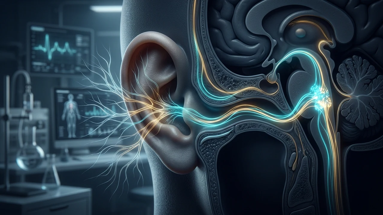
Neuroanatomy of the Pinna: The Four Neural Innervations
The external ear is a complex sensory mosaic innervated by cranial and somatic nerves. Groundbreaking micro-dissection research conducted by Peuker and Filler established the precise boundary lines between these overlapping sensory fields:
- Auricular Branch of the Vagus Nerve (ABVN / Arnold's Nerve): Exits the jugular fossa, traverses the mastoid canaliculus, and enters the external auditory canal and conchal bowl. It carries myelinated A-beta afferents that terminate directly in the medullary brainstem.
- Auriculotemporal Nerve (Cranial Nerve V3): Branch of the mandibular division of the trigeminal nerve. Innervates the anterior upper ear, anterior canal wall, and superior aspect of the tragus.
- Great Auricular Nerve (Cervical Plexus C2-C3): Supplies somatic sensory fibers to the lower half of the external ear, the entire earlobe (lobule), and the posterior retroauricular skin.
- Lesser Occipital Nerve (Cervical Plexus C2): Innervates the posterior superior margin of the helix and cranial surface of the pinna.
Cymba Conchae vs. Tragus: A Quantitative Comparison
Two primary anatomical landmarks are utilized in clinical taVNS literature: the Cymba Conchae and the Tragus. Understanding the morphological differences between these two zones is essential for optimizing current density and clinical efficacy.
| Parameter | Cymba Conchae | Tragus (Anterior & Posterior) |
|---|---|---|
| ABVN Fiber Density | 100% Exclusive Vagal Innervation | 45% - 60% Vagal (Mixed with CN V3) |
| fMRI BOLD Signal | Robust activation of NTS, Locus Coeruleus, and Parabrachial Area | Strong NTS activation, with co-activation of trigeminal sensory nuclei |
| Electrode Interface | Custom silicone/metal probe or curved cup electrode | Spring-loaded or silicone clip electrode |
| Mechanical Stability | Requires custom ear mold or earphone chassis | High; readily clamped across cartilage |
| Skin Impedance | Moderate (prone to cerumen/oil accumulation) | Low to moderate (thin cutaneous barrier) |
| Sensory Threshold (mA) | Lower sensory threshold (0.6 - 1.8 mA) | Higher sensory threshold (1.2 - 2.8 mA) |
The Cymba Conchae: The 100% Pure Vagal Harbor
The concha of the ear is anatomically divided into two chambers by the crus of the helix: the superior cavity is the cymba conchae, while the inferior cavity is the cavum conchae. The cymba conchae forms a smooth, concave shell superior to the auditory meatus.
Peuker and Filler's anatomical dissections confirmed that the cymba conchae receives 100% pure innervation from the ABVN without sensory overlap from the trigeminal or cervical spinal networks. In 2015, Frangos and colleagues performed blood-oxygen-level-dependent (BOLD) fMRI scans on healthy human volunteers during cymba conchae stimulation. They observed prominent, statistically robust activation of the ipsilateral Nucleus Tractus Solitarius (NTS), the Locus Coeruleus (the brain's master norepinephrine generator), the Parabrachial Nucleus, and concurrent deactivation of the central Amygdala. When the same current was delivered to the earlobe (sham), these medullary activations completely disappeared.
The Tragus: Mechanical Convenience with Mixed Afferents
The tragus is the small triangular cartilaginous flap projecting posteriorly over the opening of the ear canal. From a clinical perspective, the tragus remains the most widely utilized site for commercial consumer devices because it allows for a dual-sided clip electrode (cathode on the inner tragus, anode on the outer tragus).
While the tragus provides robust clinical efficacy in treating major depressive disorder, migraine, and inflammatory bowel flare-ups, clinicians must recognize that the anterior surface contains branches of the auriculotemporal nerve. Stimulating the tragus recruits both vagal afferents (projecting to the NTS) and trigeminal afferents (projecting to the spinal trigeminal nucleus). This dual-recruitment is therapeutically viable, but requires clinicians to titrate current based on comfort, ensuring that trigeminal activation does not induce facial twitching or jaw tension.
Step-by-Step Clinical Electrode Application Protocol
To maximize electrical charge transfer and prevent epidermal irritation, follow this four-stage preparation protocol:
- Skin De-Lipidation: The external ear produces sebum that dramatically raises cutaneous electrical impedance. Always cleanse the cymba conchae or tragus using a 70% isopropyl alcohol swab and allow it to dry completely. A clean skin surface reduces resistance from 25,000 Ohms down to under 5,000 Ohms.
- Conductive Medium: Dry metal electrodes cause micro-arcing and pinpoint burning sensations. Apply a pea-sized drop of isotonic chloride-based conductive gel (or saline-soaked foam tips) to both contact points.
- Polarity Configuration:
- For Tragus Clips: Position the active negative electrode (Cathode / Black) on the inner (medial) surface of the tragus, which has the highest density of ABVN endings. Place the positive reference electrode (Anode / Red) on the outer (lateral) surface.
- For Cymba Probes: Place both the cathode and anode within the conchal bowl, spaced 5 to 8 millimeters apart.
- Sensory Titration: Gradually increase the current amplitude from 0.0 mA in 0.1 mA increments until the patient reports a distinct, non-painful tingling sensation ("pins and needles"). Never titrate into sharp stinging or painful discomfort.
For patients and practitioners constructing home protocols, see our exhaustive guide on at-home vagus nerve stimulation guidelines and read our comprehensive review of published taVNS clinical trials and human data.