
1. What Is Sensory Motor Amnesia? The Neurological Breakdown
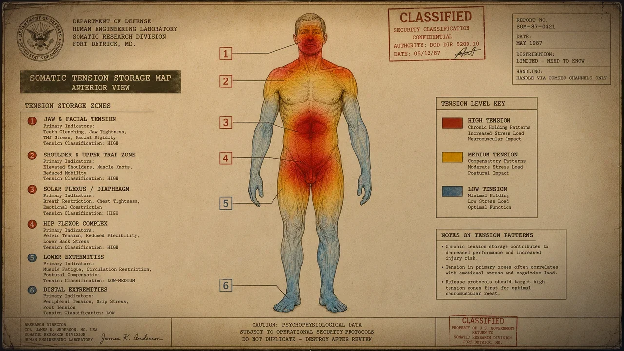
Coined in 1988 by philosopher and somatics pioneer Thomas Hanna, PhD, Sensory Motor Amnesia (SMA) describes a state in which the motor neurons of the central nervous system maintain continuous low-grade contraction in skeletal muscles, while the sensory feedback loops have habituated to the tension. The brain no longer registers that the muscle is contracted, nor does it retain voluntary neural pathways to lengthen it.
In SMA, the problem does not reside in the muscle tissue, fascia, or tendons. It is a software malfunction located in the sensorimotor cortex (precentral and postcentral gyri). Under prolonged emotional distress, repetitive postural strain, or physical trauma, subcortical reflexes take permanent command of motor output, transforming temporary defensive holding into an involuntary, unconscious baseline posture.
2. The Three Somatic Reflexes of Chronic Stress
Clinical somatics identifies three primary subcortical reflex patterns that dictate involuntary muscle contractions across the human body:
- The Red Light Reflex (Withdrawal / Startle): Triggered by fear, distress, or exhaustion. Flexes the anterior body chain: chin tucks, shoulders round, abdominal muscles clench, and hips curl forward. Chronic engagement produces shallow breathing, sunken posture, and mid-back pain.
- The Green Light Reflex (Landau / Action): Triggered by relentless productivity, deadlines, and urgency. Contracts the posterior extensor chain: lumbar erectors arch, neck hyperextends, and gluteals clench. Chronic engagement produces lumbar disc compression and sciatica.
- The Trauma Reflex (Protection / Scoliosis): Triggered by unilateral injury, surgery, or asymmetric avoidance. Rotates and tilts the pelvis and torso to shield an injured area, producing chronic hip unevenness and sacroiliac joint dysfunction.
Neuro-Somatic Insight: Gamma Motor Neuron Overdrive
In Sensory Motor Amnesia, central gamma motor efferents continually pre-set muscle spindles to a shortened baseline. Even during deep sleep or general anesthesia, electromyography (EMG) reveals residual microvolt activity in SMA-affected muscles, demonstrating that true relaxation requires sensory cortical re-education rather than physical rest.
3. Cortical Smudging: When the Brain Loses the Muscle Map
The human brain maintains a topographical representation of every body part in the sensory and motor cortices—the classic cortical homunculus. To move a muscle with precision and relax it fully, the brain requires crisp, high-resolution sensory feedback from peripheral proprioceptors.
When a muscle group is held in continuous isometric contraction for months or years, the sensory feedback becomes uniform, static, and redundant. The brain undergoes a process neuroscientists call cortical smudging: the distinct boundaries of that muscle's neural map blur into neighboring areas. When the patient is asked to isolate and relax their lower lumbar erectors or transversus abdominis, they physically cannot find the neural "switch" because the cortical map has become degraded.
4. Why Stretching and Involuntary Force Fail
The standard cultural prescription for tight muscles is passive stretching. However, from a neurophysiological standpoint, passive stretching is the least effective method for resolving SMA. When you forcefully stretch a muscle afflicted by SMA, you stimulate the myotatic stretch reflex inside the muscle spindle.
The spinal cord interprets rapid passive elongation as a threat of tissue rupture, instantaneously firing an efferent alpha motor signal that commands the muscle to contract harder. This is why aggressive yoga or static stretching frequently results in rebound soreness and muscle spasms 24 hours later. The muscle is not "short"; the nervous system is simply keeping it "switched on."
5. Somatic Exercises for Chronic Muscle Tension
To safely reverse cortical smudging and overwrite involuntary gamma firing, patients must engage in targeted somatic exercises for chronic muscle tension that exploit active motor learning.
- Somatosensory Differentiation: Moving a single joint through micro-ranges (less than 5 degrees) while mentally tracking sensations of friction, tension, and release.
- Reciprocal Inhibition Drills: Contracting the antagonist muscle group with conscious focus, which forces the spinal cord to send inhibitory signals to the spastic target muscle.
- Sub-maximal Isometric Loading: Engaging the locked muscle voluntarily at just 20% to 30% of maximum force to bring its motor units back under prefrontal conscious control.
6. The Clinical Pandiculation Protocol
The neurobiological gold standard for resolving Sensory Motor Amnesia is pandiculation—a three-step neuromuscular re-education technique observed in all vertebrate animals (such as dogs and cats yawning and stretching their spines upon waking):
- Voluntary Concentric Contraction: Actively contract the already tight muscle into a slightly tighter shortened position (to roughly 50% effort). This sends a massive burst of conscious sensory data up to the motor cortex, lighting up the smudged homunculus.
- Slow, Conscious Eccentric Lengthening: Over a span of 10 to 15 seconds, smoothly and deliberately allow the muscle to lengthen millimeter by millimeter against gentle resistance, maintaining unbroken cognitive focus on the sensation of fibers de-recruiting.
- Complete Release and Integration: Once the muscle reaches full length, pause for 5 full seconds in absolute stillness, allowing the motor cortex to register the new zero-voltage resting baseline.