
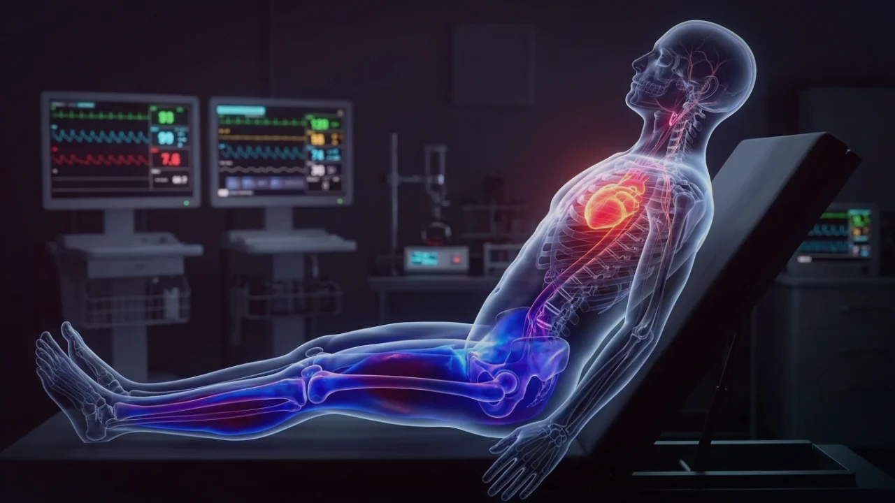
1. Defining Post-Exertional Malaise: The Cardinal Sign of Neuro-Metabolic Disease
Post-Exertional Malaise (PEM) is the cardinal diagnostic feature of Myalgic Encephalomyelitis / Chronic Fatigue Syndrome (ME/CFS) and a major component of post-viral syndromes, including Long COVID neuro-dysautonomia. The Institute of Medicine (IOM / National Academy of Medicine) defines PEM as a complex, multi-system exacerbation of symptoms following previously tolerated physical, cognitive, sensory, or emotional exertion.
Unlike normal exercise-induced fatigue, which manifests during or immediately following physical effort and resolves with standard rest, PEM is characterized by a distinctive temporal latency: symptoms typically peak 24 to 72 hours after the triggering event and can persist for days, weeks, or months. During a crash, patients suffer profound systemic weakness, orthostatic intolerance, severe cognitive slowing ("brain fog"), flu-like myalgias, lymphadenopathy, and complete autonomic instability.
2. The 2-Day Cardiopulmonary Exercise Test (CPET): Objective Metabolic Proof
For decades, skeptical clinicians dismissed PEM as subjective deconditioning, psychosomatic illness, or kinesiophobia. This controversy was decisively resolved through the implementation of the 2-day Cardiopulmonary Exercise Testing (CPET) protocol, spearheaded by researchers at the Workwell Foundation and Pacific University.
A standard single CPET can establish baseline peak oxygen consumption ($VO_2peak$), but it cannot evaluate cellular recovery capacity. In healthy sedentary individuals, elite athletes, and even patients with severe chronic heart failure or COPD, performing a maximal CPET on Day 1 does not impair performance on an identical CPET repeated 24 hours later (Day 2); in fact, performance parameters are reproducible to within ±5% due to familiarization.
In patients with PEM, however, Day 2 testing reveals an unprecedented metabolic collapse. On Day 2, patients show:
- A 15% to 30% reduction in $VO_2$ at the anaerobic (ventilatory) threshold ($VO_2$ at AT).
- A dramatic drop in peak workload (measured in Watts) achieved.
- Inability to reach predicted peak heart rates due to chronotropic incompetence.
- Severe worsening of ventilatory efficiency ($V_E/VCO_2$ slope).
This failure to reproduce physiological baseline work is unique in exercise physiology and provides indisputable objective biomarker proof of acute bioenergetic and mitochondrial dysfunction.
3. Biochemical Mechanisms: Oxygen Extraction Deficits & Lactic Acid Spikes
What occurs at the cellular level during a PEM crash? Invasive cardiopulmonary exercise testing (iCPET) conducted at Brigham and Women's Hospital by David Systrom has revealed the root hemodynamic defect: impaired systemic oxygen extraction ($C(a-v)O_2$ difference).
Even though arterial oxygen saturation ($SaO_2$) is 98% to 100%, skeletal muscle myocytes and neurons fail to extract oxygen from capillary blood. Mixed venous oxygen saturation remains abnormally high, indicating that oxygen is shunting through microvascular beds without being taken up into mitochondria. Inside the myocytes, mitochondrial Complex I, Complex II, and ATP synthase are functionally impaired. Consequently, cells cannot sustain pyruvate oxidation through the Krebs cycle, forcing cells into early anaerobic glycolysis at remarkably low workloads—often at heart rates under 100 bpm—causing rapid intracellular lactic acidosis and tissue injury.
4. PEM vs. True Cardiovascular Deconditioning (Comparison Matrix)
The clear clinical and biochemical distinctions between true deconditioning and post-exertional malaise are contrasted below:
| Diagnostic Feature | Cardiovascular Deconditioning | Post-Exertional Malaise (PEM) |
|---|---|---|
| Day 2 CPET Reproducibility | 100% reproducible; VO2 at AT remains identical or improves (±5%) | Catastrophic drop: 15% to 30% collapse in VO2 and workload at AT |
| Response to Exercise Training | Progressive improvement in aerobic capacity and stamina over weeks | Severe clinical deterioration; crashes lasting days to weeks |
| Temporal Symptom Onset | Immediate fatigue during exercise; normalizes rapidly post-rest | Delayed onset; typically peaks 24 to 72 hours after exertion |
| Anaerobic Lactic Threshold | Normal for age/conditioning level; shifts upward with training | Pathologically low; patient enters lactic acidosis during light daily tasks |
| Systemic Biological Symptoms | Musculoskeletal fatigue; no immune or cognitive flare-ups | Flu-like symptoms, brain fog, lymph node pain, orthostatic tachycardia |
| Underlying Pathophysiology | Decreased cardiac stroke volume and plasma volume | Mitochondrial electron transport chain failure; microvascular shunting |
5. Microvascular Endothelial Dysfunction & Impaired Oxygen Shuttling
A secondary driver of PEM is microvascular endothelial dysfunction and autonomic dysregulation of peripheral blood distribution. In a healthy subject, sympathetic withdrawal and local endothelial nitric oxide release induce arteriolar vasodilation in exercising muscle, delivering oxygen exactly where needed.
In patients with PEM and dysautonomia, endothelial nitric oxide synthase (eNOS) uncoupling and autonomic small-fiber neuropathy prevent coordinated vasodilation. Blood is diverted through functional arteriovenous shunts, bypassing nutritive capillary beds. This results in tissue hypoxia despite normal macrovascular blood flow, triggering severe local ischemia, oxidative stress, and the release of inflammatory damage-associated molecular patterns (DAMPs) into circulation.
6. Clinical Management: Pacing, Heart Rate Gating & Metabolic Recovery
Because traditional graded exercise therapy (GET) is contraindicated and clinically harmful in PEM, management centers on pacing within the metabolic energy envelope:
- Heart Rate Biofeedback Gating (The Ventilatory Threshold Limit): Calculate the patient's estimated resting anaerobic threshold (often approximated as $[220 - ext{Age}] imes 0.55$ to $0.60$, or precisely measured via CPET). Patients wear continuous heart rate monitors with audible alarms, pausing immediately whenever heart rate approaches this threshold to prevent entering anaerobic glycolysis.
- Cognitive and Sensory Pacing: Because intensive cognitive tasks (reading, screens, emotional conversations) consume substantial cerebral glucose and ATP, cognitive exertion must be broken into 15-to-20-minute intervals interspersed with radical rest (lying flat in a quiet, dark room).
- Orthostatic Load Reduction: In patients with comorbid POTS, performing physical movements in recumbent or semi-recumbent positions (supine stretching, floor yoga, recumbent cycling) eliminates the gravity-induced orthostatic sympathetic surge, saving valuable metabolic ATP for vital cellular repair.