
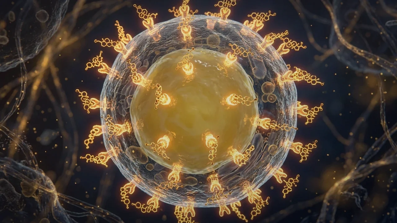
1. Biochemical Architecture: Subcellular NAD+ Compartmentation
Nicotinamide adenine dinucleotide exists in two distinct functional states: the oxidized form ($NAD^+$) and the reduced form ($NADH$). While the ratio of $NAD^+/NADH$ governs the thermodynamics of glycolysis and the tricarboxylic acid (TCA) cycle, the absolute concentration of free $NAD^+$ acts as a limiting regulatory ligand for non-redox signaling enzymes.
Within mammalian neurons, NAD+ is compartmentalized into three distinct subcellular pools: cytosolic, nuclear, and mitochondrial. Because the inner mitochondrial membrane is impermeable to intact pyridine nucleotides, mitochondria possess a dedicated, recently discovered mammalian NAD+ carrier (SLC25A51). While cytosolic and nuclear NAD+ levels can fluctuate rapidly in response to metabolic stress, the mitochondrial pool is buffered with high priority, often maintaining concentrations of 250 to 500 $mu M$ to sustain basal oxidative phosphorylation.
In neurons, de novo synthesis of NAD+ from dietary tryptophan via the kynurenine pathway is negligible. Instead, greater than 99% of neuronal NAD+ is maintained through the salvage pathway, where nicotinamide ($NAM$) is recycled back to nicotinamide mononucleotide ($NMN$) by the rate-limiting enzyme nicotinamide phosphoribosyltransferase (NAMPT), followed by adenylation to NAD+ by NMNAT1-3.
2. Sirtuin Enzymology: SIRT1, SIRT3 & Respiratory Chain Activation
Sirtuins are evolutionary conserved class III histone deacetylases that have an obligate requirement for $NAD^+$ to cleave acetyl groups from lysine residues on target proteins, generating nicotinamide and 2'-O-acetyl-ADP-ribose. In the central nervous system, two sirtuins dictate mitochondrial integrity:
- SIRT1 (Nuclear / Cytosolic): Deacetylates the master transcriptional coactivator PGC-1α, promoting its nuclear translocation and driving the transcription of nuclear-encoded mitochondrial genes (NRF-1, NRF-2, and TFAM), effectively stimulating mitochondrial biogenesis.
- SIRT3 (Mitochondrial Matrix): The primary protein deacetylase within mitochondria. In the presence of adequate NAD+, SIRT3 deacetylates NDUFA9 of Complex I and the catalytic subunit of ATP synthase (Complex V), boosting electron flux and ATP generation. Furthermore, SIRT3 deacetylates Superoxide Dismutase 2 (MnSOD) and isocitrate dehydrogenase 2 (IDH2), elevating the mitochondrial antioxidant defense capacity to neutralize reactive oxygen species.
When cellular NAD+ concentrations drop below the $K_m$ of these enzymes (~100–200 $mu M$ for SIRT1/3), sirtuin enzymatic velocity plummets. Mitochondrial proteins accumulate excessive acetyl adducts, leading to structural instability of electron transport chain supercomplexes and catastrophic bioenergetic failure.
3. The Competitive Sinks: CD38 Upregulation & PARP Hyperactivation
Why do neuronal and glial NAD+ pools deplete during chronic physiological duress? The collapse is driven by competitive over-activation of two major NAD+-consuming enzyme families:
- Poly(ADP-ribose) Polymerases (PARPs, primarily PARP-1): Activated by single- and double-strand DNA breaks caused by oxidative stress and lipid peroxidation. Upon activation, PARP-1 consumes massive amounts of NAD+ to synthesize branched poly(ADP-ribose) polymers on nuclear histones to recruit DNA repair complexes. In states of persistent oxidative stress, PARP-1 becomes hyperactivated, draining cellular NAD+ faster than the NAMPT salvage pathway can regenerate it.
- CD38 (Cyclic ADP-Ribose Hydrolase): A type II transmembrane glycoprotein expressed on astrocytes, microglia, and endothelial cells. CD38 is an exceptionally inefficient enzyme, hydrolyzing approximately 100 molecules of NAD+ for every single molecule of cyclic ADP-ribose produced. Pro-inflammatory cytokines ($TNF-alpha$, $IL-6$, $IFN-gamma$) dramatically upregulate CD38 expression, turning it into a massive "metabolic sink" that destroys tissue NAD+ reserves.
4. Sirtuin Family Isoforms in Neuro-Autonomic Function (Comparison Matrix)
The differential tissue localization, enzymatic targets, and physiological functions of the human sirtuin family are outlined below:
| Sirtuin Isoform | Subcellular Location | NAD+ Sensitivity ($K_m$) | Primary Molecular Targets | Autonomic & Neurological Role |
|---|---|---|---|---|
| SIRT1 | Nucleus & Cytosol | Moderate (~150–200 μM) | PGC-1α, FOXO3a, NF-κB (p65), p53 | Stimulates mitochondrial biogenesis; suppresses microglial neuroinflammation |
| SIRT2 | Cytosol & Nucleus | High (~200–300 μM) | Alpha-tubulin, FOXO1, PEPCK | Regulates myelin sheath maintenance and axonal structural stability |
| SIRT3 | Mitochondrial Matrix | High (~100–250 μM) | Complex I, Succinate Dehydrogenase, MnSOD | Master mitochondrial engine: Maximizes ATP output and antioxidant defense |
| SIRT4 | Mitochondrial Matrix | Very High (ADP-ribosyltransferase) | Glutamate Dehydrogenase (GDH), ANT | Regulates amino acid-stimulated insulin secretion and mitochondrial glutamine metabolism |
| SIRT5 | Mitochondrial Matrix | Demalonylase / Desuccinylase | CPS1, Cytochrome c, Succinate DH | Eliminates acidic dicarboxylic acyl lysine modifications; protects against oxidative shock |
5. Clinical Manifestations: Neuro-Metabolic Exhaustion & Dysautonomia
When brainstem autonomic centers—including the dorsal motor vagal nucleus and the rostral ventrolateral medulla—suffer from NAD+ depletion, the clinical phenotype is characterized by autonomic brittleness. Pacemaker neurons in these nuclei lose their ability to sustain rapid firing without triggering energy failure.
Patients present with profound, unyielding physical exhaustion that does not improve with rest, orthostatic heart rate spikes (POTS), sudden drops in baroreflex sensitivity, and marked exercise intolerance. Furthermore, because SIRT1 inhibition relieves suppression on the NF-κB transcription factor, central neuroinflammation escalates, driving severe brain fog, sensory overload, and sleep fragmentation.
6. Evidence-Based Protocols for Restoring Cellular NAD+ Flux
Re-establishing optimal NAD+ levels and sirtuin signaling requires a multi-target approach that boosts synthesis while blocking uncontrolled degradation:
- Precursor Replenishment (NMN / NR): Supplementing with bioavailable salvage pathway precursors—such as Nicotinamide Mononucleotide (NMN) or Nicotinamide Riboside (NR)—bypasses the rate-limiting NAMPT enzyme, directly elevating intracellular NAD+ concentrations and restoring SIRT1/3 activity.
- Inhibition of Pathological CD38 Activity: Naturally occurring flavonoids, specifically Apigenin (derived from chamomile and celery) and Quercetin, act as potent, cell-permeable competitive inhibitors of the CD38 ecto-enzyme, cutting NAD+ waste by up to 50% in inflamed tissues.
- Allosteric Sirtuin Activation (Resveratrol / Pterostilbene): Polyphenolic sirtuin-activating compounds (STACs) bind directly to SIRT1, inducing a conformational shift that increases its binding affinity for NAD+ and acetylated peptide substrates, amplifying mitochondrial biogenesis even in the presence of moderate NAD+ limitations.