
1. The High-Energy Demands of Central Autonomic Circuits
Neurons are post-mitotic cells characterized by extensive dendritic arborizations and elongated axonal projections that extend across substantial anatomical distances. To propagate action potentials and mediate chemical neurotransmission, a neuron must continuously pump sodium ($Na^+$) out of the cytoplasm and potassium ($K^+$) into the intracellular compartment against steep electrochemical gradients. This task is governed by the $Na^+/K^+$-ATPase enzyme, which hydrolyzes a single ATP molecule for every three $Na^+$ ions expelled and two $K^+$ ions imported.
Within the central autonomic network (CAN)—including the solitary tract nucleus (NTS), the rostral ventrolateral medulla (RVLM), the hypothalamus, and the dorsal motor nucleus of the vagus (DMV)—neurons fire tonically without pause throughout life to regulate baroreflex sensitivity, vascular tone, and cardiorespiratory rhythms. Consequently, these autonomic pacemaker centers have the highest density of mitochondria per unit volume of any biological tissue. Any drop in mitochondrial ATP synthesis directly impairs autonomic repolarization, causing instability in blood pressure regulation, aberrant heart rate swings, and severe central exhaustion.
2. Electron Transport Chain Decoupling & Reactive Oxygen Species
ATP is generated through oxidative phosphorylation across the inner mitochondrial membrane via five multi-protein complexes (Complexes I through V). Reducing equivalents ($NADH$ and $FADH_2$) derived from the tricarboxylic acid (TCA) cycle donate high-energy electrons, which pass along the chain to molecular oxygen, while protons ($H^+$) are pumped into the intermembrane space to establish an electrochemical proton-motive force.
Under conditions of chronic inflammatory signaling (such as persistent $TNF-\alpha$ and $IL-1\beta$ exposure), electron transfer kinetics stall, primarily at Complex I (NADH:ubiquinone oxidoreductase) and Complex III (cytochrome bc1 complex). When electron flow is retarded, premature single-electron transfers to oxygen occur, generating superoxide anions ($O_2^{\bullet-}$). Superoxide dismutates into hydrogen peroxide ($H_2O_2$), which in the presence of trace transition metals undergoes Fenton chemistry to produce hydroxyl radicals ($^{\bullet}OH$)—the most destructive reactive oxygen species known to cellular biology.
Hydroxyl radicals attack the polyunsaturated fatty acids within cardiolipin, a specialized phospholipid essential for maintaining mitochondrial inner membrane curvature and stabilizing respiratory supercomplexes. Cardiolipin peroxidation leads to cristae unraveling, release of cytochrome c into the cytosol, and profound collapse of the trans-membrane electrical potential ($DeltaPsi_m$), rendering ATP synthase functionally paralyzed.
3. Mitochondrial Dynamics: Fission, Fusion & Mitophagy in Neurons
Mitochondria in healthy neurons do not operate as static, isolated entities; they exist as dynamic, constantly remodeling reticular networks regulated by balanced cycles of mitochondrial fusion (promoted by Mitofusins Mfn1/Mfn2 and OPA1) and mitochondrial fission (driven by Dynamin-Related Protein 1, Drp1).
Fusion allows damaged mitochondria to intermix contents with healthy organelles, diluting mutated mtDNA and replenishing depleted antioxidant proteins. Fission, conversely, isolates terminally damaged, depolarized segments of the mitochondrial network for degradation via receptor-mediated autophagy, termed mitophagy (orchestrated by the PINK1/Parkin kinase pathway). Under sustained allostatic load and elevated glucocorticoid levels, the balance shifts pathologically toward hyper-fission. Fragmented, swollen mitochondria accumulate in axon terminals, incapable of sustaining local synaptic ATP supply while generating excessive oxidative stress.
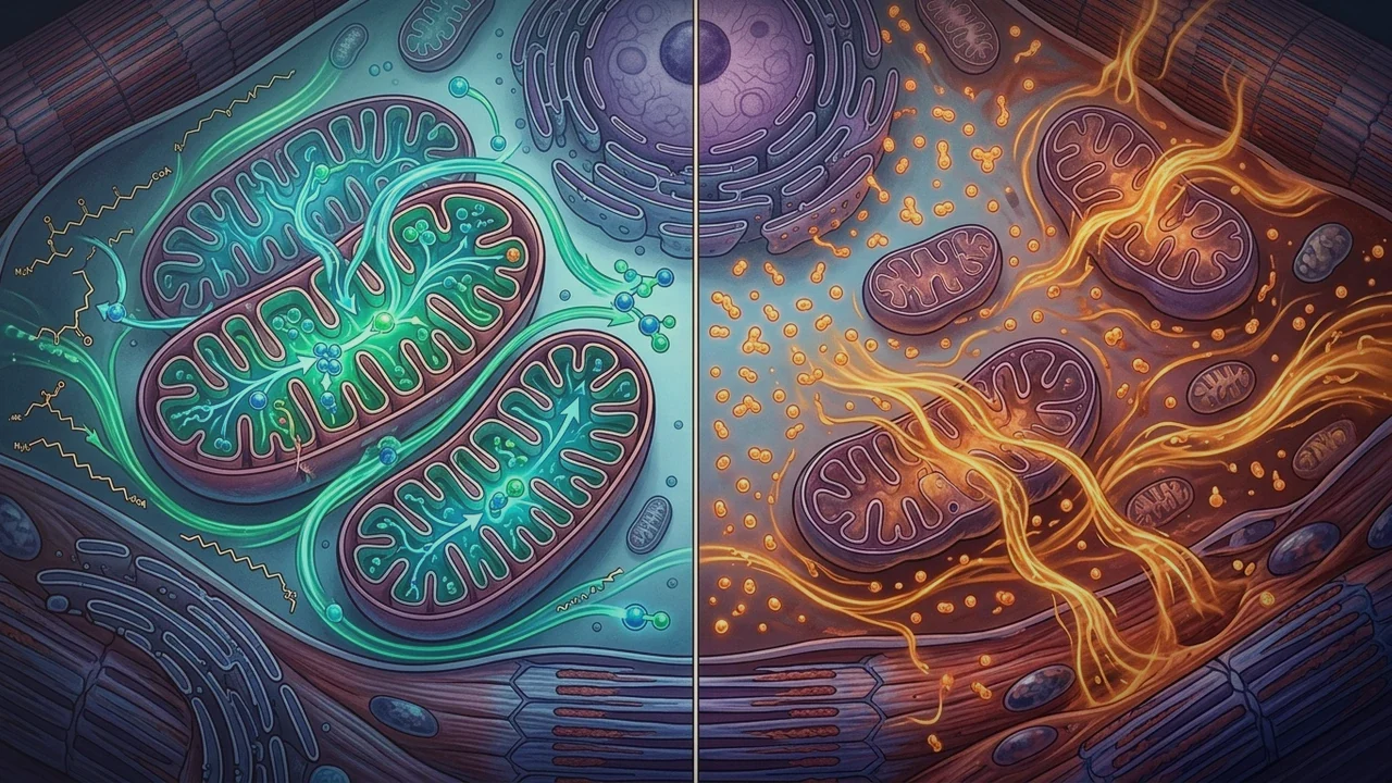
4. Oxidative Phosphorylation vs. Aerobic Glycolysis (Comparison Matrix)
When mitochondrial respiratory chains fail, neural tissue attempts to compensate through anaerobic and aerobic glycolytic pathways, resulting in profound metabolic tradeoffs:
| Metabolic Feature | Healthy Oxidative Phosphorylation | Mitochondrial Failure / Glycolytic Shift |
|---|---|---|
| ATP Yield per Glucose Molecule | 30 to 32 ATP molecules (high efficiency) | 2 net ATP molecules (low efficiency, 15x less energy) |
| Oxygen Dependence | Strictly obligate ($O_2$ terminal electron acceptor) | Anaerobic or oxygen-independent (Warburg effect) |
| Metabolic End-Product | Water ($H_2O$) and Carbon Dioxide ($CO_2$) | Lactate ($C_3H_5O_3^-$) and Protons ($H^+$) |
| Impact on Tissue Microenvironment | Neutral pH, physiological carbon dioxide tension | Local tissue acidosis, microglial activation, vascular spasm |
| Substrate Flexibility | Glucose, Ketones (Beta-hydroxybutyrate), Lactate | Exclusively dependent on rapid extracellular glucose uptake |
| Clinical Manifestation | Sustained mental clarity, stable autonomic tone | Profound post-exertional crash, lactic burn, brain fog |
5. Clinical Manifestations: Dysautonomia, POTS & Post-Exertional Fatigue
The clinical hallmark of neuronal mitochondrial dysfunction is post-exertional malaise (PEM) and autonomic instability. Because sympathetic ganglia and parasympathetic vagal motor nuclei require continuous ATP to synthesize acetylcholine and noradrenaline, bioenergetic depletion precipitates immediate failure of autonomic reflex buffering.
Upon standing, a healthy individual's sympathetic nervous system releases norepinephrine at mesenteric and splanchnic vascular beds to induce vasoconstriction. In the presence of mitochondrial ATP exhaustion, vascular smooth muscle cells and sympathetic postganglionic axons fail to sustain vasoconstrictive tone. Gravitational venous pooling ensues, triggering compensatory sinus tachycardia (POTS) and cerebral hypoperfusion, presenting clinically as orthostatic dizziness, palpitations, and pervasive cognitive exhaustion.
6. Evidence-Based Metabolic Strategies for Mitochondrial Restoration
Reversing mitochondrial exhaustion requires stimulating mitochondrial biogenesis, optimizing substrate delivery, and restoring the redox environment:
- Activation of the PGC-1α Pathway: Peroxisome proliferator-activated receptor-gamma coactivator 1-alpha is the master transcriptional regulator of mitochondrial biogenesis. Low-intensity, steady-state Zone 2 aerobic training (when tolerated without triggering PEM) stimulates AMP-activated protein kinase (AMPK) and Sirtuin-1 (SIRT1), driving PGC-1α nuclear translocation.
- Restoring NAD+ Availability: Nicotinamide adenine dinucleotide ($NAD^+$) is an essential cofactor for mitochondrial sirtuins (SIRT3/4/5) that deacetylate and activate electron transport chain complexes. Supporting NAD+ salvage pathways preserves mitochondrial enzymatic activity under oxidative duress.
- Cofactor Replenishment: Targeted cofactors—including Ubiquinol (active CoQ10), Alpha-Lipoic Acid (ALA), Acetyl-L-Carnitine, and Magnesium Bisglycinate—serve as electron shuttles and fatty acid transport carriers, stabilizing inner mitochondrial membrane potentials.