
1. Neurophysiology of Glutamate: Receptors, Synapses & Homeostasis
Glutamate ($L-glutamate$) is the primary excitatory amino acid in the brain. Because extracellular glutamate concentrations in excess of 1 to 2 micromolar ($mu M$) are cytotoxic to neurons, synaptic levels are maintained within strict spatial and temporal boundaries. Upon depolarization, presynaptic vesicles release glutamate into the synaptic cleft, where it reaches peak concentrations of approximately 1 millimolar ($mM$) for only a few milliseconds before being cleared.
This rapid clearance is performed not by enzymatic degradation in the cleft, but by high-affinity sodium-dependent transporters: Excitatory Amino Acid Transporters (EAAT1 and EAAT2), located primarily on the membranes of surrounding perisynaptic astrocytic processes. Inside the astrocyte, glutamate is converted into inert glutamine by the enzyme glutamine synthetase, shuttled back into the presynaptic neuron, and hydrolyzed back into glutamate by glutaminase—a closed loop known as the glutamate-glutamine cycle.
2. Molecular Mechanics: The Magnesium Block & Pathological NMDA Gating
Postsynaptic glutamate signaling operates through ionotropic receptors (AMPA, Kainate, and NMDA) and metabotropic receptors (mGluR1-8). The NMDA receptor is a heterotetrameric ion channel composed of GluN1 and GluN2 subunits that is uniquely voltage-dependent and highly permeable to calcium ($Ca^{2+}$).
Under resting physiological membrane potentials (-70 mV), an extracellular hydrated magnesium ion ($Mg^{2+}$) lodges within the channel pore, physically blocking ion conductance even when glutamate and its co-agonist glycine are bound. Only when adjacent AMPA receptors depolarize the membrane to approximately -30 mV is the $Mg^{2+}$ ion electrostatically expelled, allowing calcium to enter.
In states of chronic cellular stress, low ATP availability, or severe systemic magnesium deficiency, resting membrane potential drifts upward, and the protective magnesium block is lost. The NMDA receptor remains continuously gated open, transforming an informative synaptic pulse into a chronic, lethal calcium inundation.
3. The Calcium Cascade: Calpains, NOS Activation & Mitochondrial Collapse
Once cytosolic calcium surges beyond the buffering capacity of the endoplasmic reticulum and calbindin proteins, a catastrophic enzymatic death cascade is initiated:
- Calpain & Phospholipase Activation: Excessive $Ca^{2+}$ activates calpain, a calcium-dependent neutral cysteine protease that hydrolyzes the neuronal cytoskeleton (spectrin, MAP2) and degrades EAAT2 transporters. Simultaneously, phospholipase A2 ($PLA_2$) is unleashed, cleaving membrane phospholipids into arachidonic acid and inflammatory leukotrienes.
- Neuronal Nitric Oxide Synthase (nNOS): Calcium activates nNOS to produce massive amounts of nitric oxide ($NO$). Nitric oxide reacts with superoxide ($O_2^{\bullet-}$) to form peroxynitrite ($ONOO^-$)—a highly reactive nitrogen species that nitrates tyrosine residues, irreversibly inhibiting Complex I of the mitochondrial electron transport chain.
- Mitochondrial Permeability Transition Pore (mPTP): Mitochondria avidly sequester cytosolic calcium until their matrix capacity is overwhelmed. High intramitochondrial calcium triggers the opening of the large-conductance mPTP, collapsing the inner membrane proton gradient, arresting ATP synthesis, and releasing pro-apoptotic factors (cytochrome c and apoptosis-inducing factor) into the cytosol.
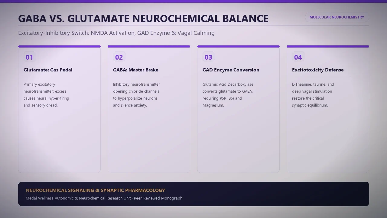
4. Glutamate Excitation vs. GABAergic Inhibition (Comparison Matrix)
The neurochemical balance between excitation and inhibition dictates autonomic stability and subjective calm:
| Neurochemical Parameter | Glutamate (Excitatory Axis) | GABA (Inhibitory Axis) |
|---|---|---|
| Primary Receptors | NMDA, AMPA, Kainate, mGluR1–8 | GABA-A (ligand-gated chloride channel), GABA-B (G-protein coupled) |
| Ion Permeability | Sodium ($Na^+$), Potassium ($K^+$), High Calcium ($Ca^{2+}$) | Chloride ($Cl^-$) influx → membrane hyperpolarization |
| Enzymatic Synthesis | Synthesized from glutamine via glutaminase | Synthesized from glutamate via Glutamate Decarboxylase (GAD65/67 + B6) |
| Impact on Membrane Potential | Depolarization (brings neuron closer to action potential threshold) | Hyperpolarization (drives membrane potential further from threshold) |
| Excess / Pathological State | Excitotoxicity, seizures, severe panic, sensory hypersensitivity | Sedation, ataxia, respiratory depression, blunted cognition |
| Deficiency State | Cognitive dulling, impaired synaptic plasticity, memory deficits | Intractable anxiety, muscle twitches, hyperacusis, insomnia |
5. Clinical Manifestations: Neuro-Agitation, Sensory Flooding & Dysautonomia
In clinical practice, patients with chronic NMDA hyperactivation rarely present with focal neurological deficits; instead, they suffer from an agonizing syndrome of cellular neuro-agitation. Sensory processing centers in the temporal and parietal cortices become hypersensitized, leading to hyperacusis (inability to tolerate normal sounds) and photophobia (painful sensitivity to indoor lighting).
Crucially, because the basolateral amygdala and hypothalamic paraventricular nucleus rely on glutamatergic drive to trigger sympathetic fight-or-flight outflow, excitotoxic excess locks the autonomic nervous system into permanent sympathetic overdrive, driving benign muscle fasciculations, resting tachycardia, nocturnal panic attacks, and severe somatic dread that fails to respond to cognitive reappraisal techniques.
6. Evidence-Based Protocols to Buffer Excitotoxicity and Restore Balance
Mitigating glutamate excitotoxicity requires restoring astrocytic transporter function, replenishing NMDA channel blockers, and optimizing GABA synthesis:
- Physiological NMDA Pore Blockade (Magnesium L-Threonate): Unlike standard magnesium oxide or citrate which exhibit negligible blood-brain barrier penetration, magnesium L-threonate efficiently raises cerebrospinal fluid magnesium concentrations, reinstating the voltage-dependent pore block on hyperactive NMDA channels.
- Cofactor Support for GAD Enzyme (Pyridoxal-5-Phosphate): The conversion of excitatory glutamate into inhibitory GABA is catalyzed by glutamate decarboxylase (GAD), which has an obligate requirement for active Vitamin B6 (P5P). Supplementing with bioavailable P5P prevents the accumulation of unbuffered glutamate precursors.
- Upregulation of Astrocytic EAAT2 Clearance: The amino acid derivative N-Acetylcysteine (NAC) modulates the cystine-glutamate antiporter (system $x_c^-$) and upregulates astrocytic EAAT2 expression through anti-inflammatory mechanisms, accelerating synaptic glutamate clearance.