
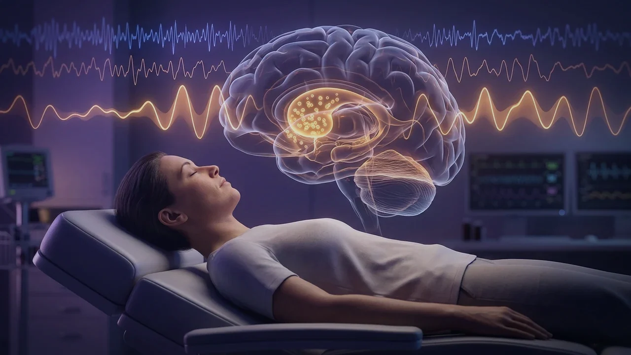
1. Pineal Neurobiology: Tryptophan to Melatonin & Ventricular Secretion
Melatonin ($N-acetyl-5-methoxytryptamine$) is an evolutionary ancient indoleamine synthesized from the essential amino acid L-tryptophan. In the pinealocyte, tryptophan is converted to serotonin ($5-HT$) via tryptophan hydroxylase and aromatic L-amino acid decarboxylase. During darkness, sympathetic postganglionic fibers from the superior cervical ganglion release norepinephrine onto pineal beta-1 and alpha-1 adrenergic receptors, dramatically activating the rate-limiting enzyme Serotonin N-acetyltransferase (AANAT).
AANAT converts serotonin into N-acetylserotonin, which is methylated by acetylserotonin O-methyltransferase (ASMT) to form lipophilic melatonin. In addition to releasing melatonin into adjacent fenestrated capillaries for systemic distribution, the pineal gland secretes melatonin directly into the cerebrospinal fluid of the third cerebral ventricle via the pineal recess. CSF melatonin concentrations reach levels 10-to-20-fold higher than plasma levels, directly bathing adjacent hypothalamic neuroendocrine centers (including the SCN and PVN) with high-potency neuroprotective signaling.
2. Receptor Pharmacology: MT1 vs. MT2 Signaling Cascades
Melatonin exerts its physiological actions via two high-affinity seven-transmembrane G-protein-coupled receptors:
- The MT1 Receptor ($Mel_{1a}$): Coupled predominantly to pertussis toxin-sensitive $G_i/G_o$ proteins. Activation of MT1 inhibits adenylyl cyclase, decreasing cyclic AMP ($cAMP$) and protein kinase A ($PKA$) activity, while opening G-protein-coupled inwardly rectifying potassium ($GIRK$) channels. In the suprachiasmatic nucleus (SCN), MT1 activation hyperpolarizes pacemaking neurons, acutely suppressing metabolic firing and driving sleep initiation.
- The MT2 Receptor ($Mel_{1b}$): Coupled to both $G_i$ and $G_q$ proteins, inhibiting soluble guanylyl cyclase and modulating intracellular calcium. In the SCN, MT2 activation triggers protein kinase C ($PKC$) pathways that induce circadian phase shifts (phase advance or phase delay), synchronizing the timing of the molecular clock with environmental night.
3. The Mitochondrial Antioxidant Engine: Scavenging Hydroxyl Radicals
Remarkably, accumulating evidence confirms that melatonin is also synthesized endogenously within the mitochondria of non-pineal cells (including neurons, astrocytes, and cardiomyocytes). Mitochondria actively uptake circulating melatonin from the cytoplasm via PEPT1 and PEPT2 oligopeptide transporters, concentrating melatonin in the matrix at levels 100 times higher than in blood plasma.
Inside the mitochondrion, melatonin acts as a direct, unmediated scavenger of the lethal hydroxyl radical ($^{\bullet}OH$), hydrogen peroxide ($H_2O_2$), and peroxynitrite ($ONOO^-$). Unlike classical antioxidants (such as Vitamin C or Vitamin E), which undergo a single redox cycle and can become pro-oxidant radicals themselves, melatonin undergoes a free radical scavenging cascade: when melatonin reacts with a radical, it transforms into cyclic 3-hydroxymelatonin, which further scavenges radicals, transforming sequentially into AFMK and AMFK. Through this cascade, a single melatonin molecule can neutralize up to 10 reactive oxygen species, protecting inner membrane cardiolipin and Complex I from oxidative destruction.
4. Melatonin Receptor Subtypes (MT1 vs. MT2 Comparison Matrix)
A detailed comparison of the pharmacological, biochemical, and physiological differences between MT1 and MT2 receptors is summarized below:
| Pharmacological Parameter | MT1 Receptor (Mel1a) | MT2 Receptor (Mel1b) |
|---|---|---|
| Primary G-Protein Coupling | $G_i / G_o$ (Inhibits adenylyl cyclase, lowers cAMP) | $G_i / G_q$ (Inhibits adenylyl & guanylyl cyclase, modulates PKC) |
| Affinity for Melatonin ($K_i$) | Very High affinity (~0.1 nM) | High affinity (~0.5–1 nM) |
| Primary SCN Function | Suppresses neuronal firing; drives acute sleep onset | Regulates circadian phase resetting and entrainment |
| Vascular Hemodynamic Effect | Mediates cerebral and peripheral vasoconstriction / stabilization | Mediates peripheral vasodilation (coronary and femoral vessels) |
| Immune & Microglial Action | Suppresses pro-inflammatory cytokine production via NF-κB | Promotes anti-inflammatory leukocyte adherence modulation |
| Synthetic Selective Agonists | Ramelteon (dual MT1/MT2), Tasimelteon | IIK-7 (selective MT2 agonist) |
5. Autonomic Remodeling: Central Vagal Stimulation & Sympathetic Quenching
Beyond its somnogenic effects, nocturnal melatonin exerts profound, direct remodeling actions on the autonomic nervous system. Melatonin receptors are densely expressed in brainstem autonomic command centers, including the rostral ventrolateral medulla (RVLM) and the nucleus ambiguus.
In the RVLM, MT1 signaling inhibits presynaptic glutamate release, dampening sympathetic vasomotor drive to the heart and kidneys and lowering systemic vascular resistance. Simultaneously, in the nucleus ambiguus, melatonin enhances cardioinhibitory vagal efferent firing. In clinical human trials, physiological nocturnal melatonin administration increases high-frequency heart rate variability (HF-HRV), restores the normal 10% to 20% nocturnal blood pressure and heart rate dip, and reduces nocturnal arrhythmias.
6. Clinical Chronotherapy Protocols for Optimizing Endogenous Melatonin
To capture the systemic and autonomic benefits of melatonin without suppressing natural endogenous synthesis, utilize targeted chronobiological protocols:
- Evening Blue-Light Blockade: Melanopsin-expressing ipRGCs suppress pineal AANAT enzyme transcription in response to wavelengths between 460 and 480 nm. Wearing amber-tinted blue-blocking eyewear or utilizing incandescent/red lighting 2 hours before bed preserves the natural onset of the dim-light melatonin surge (DLMO).
- Micro-Dose Exogenous Supplementation (0.3 mg to 1.0 mg): When supplementing, avoid supraphysiological commercial doses (5 mg to 10 mg), which desensitize MT1 and MT2 receptors and disrupt daytime circadian sensitivity. Micro-doses of 300 $mu g$ to 1 mg taken 60 to 90 minutes before sleep replicate natural peak physiological plasma levels (100–200 pg/mL) without receptor downregulation.
- Thermal Coordination: Because melatonin acts synergistically with core body temperature cooling to induce peripheral vasodilation, maintaining a cold sleeping environment (65–68°F / 18–20°C) optimizes MT2-mediated vascular response.