
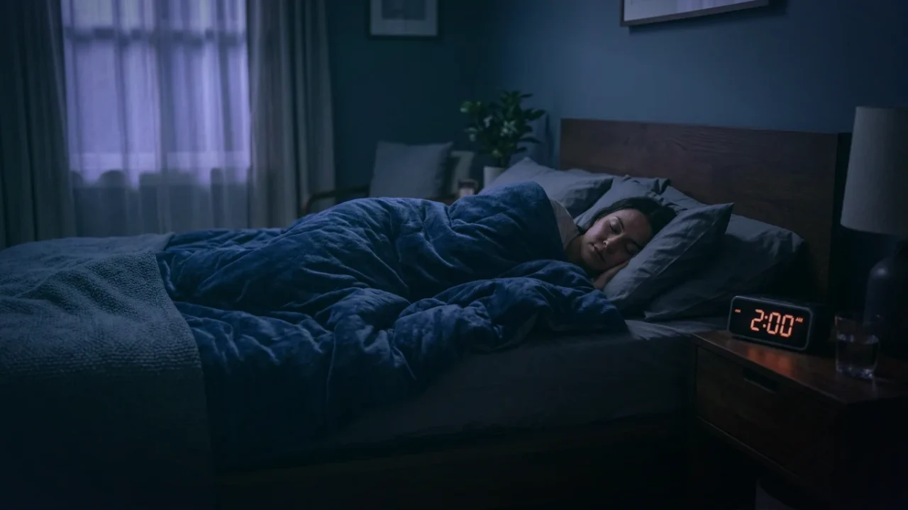
1. Biomechanics of Recumbent Body Posture and Cerebral Fluid Dynamics
The human brain is suspended within the rigid cranium, floating in approximately 150 mL of cerebrospinal fluid (CSF). The physical movement of this fluid through perivascular conduits and across the dense parenchymal extracellular matrix is governed by the laws of hydrostatic pressure, vascular pulsatility, and gravity.
When an individual transitions from an upright to a recumbent posture, the hydrostatic column changes immediately. Central venous pressure rises, intracranial venous sinuses engorge, and carotid-vertebral arterial hemodynamics adapt. However, the precise orientation of the body during recumbency—whether rotated onto the lateral axis, flat on the dorsum (supine), or ventral (prone)—imposes distinct mechanical constraints on cerebral venous drainage and perivascular space dimensions.
2. DCE-MRI Evidence: Quantifying Glymphatic Influx in Different Postures
The pivotal investigation linking body posture to glymphatic function was published in The Journal of Neuroscience by Hedok Lee, Helene Benveniste, and Maiken Nedergaard. Utilizing dynamic contrast-enhanced magnetic resonance imaging (DCE-MRI) alongside kinetic modeling in anesthetized subjects, the researchers tracked the parenchymal penetration of the paramagnetic contrast agent gadolinium-diethylenetriaminepentaacetic acid (Gd-DTPA).
The findings were striking: glymphatic transport was markedly more efficient in the lateral position compared to supine or prone positions. Microdialysis measurements of endogenous amyloid-beta and optical imaging confirmed that tracer clearance was significantly higher when animals were positioned laterally. Interestingly, ethological studies observe that the vast majority of wild mammals and humans naturally spend the preponderance of their sleep cycles in lateral recumbency, suggesting an evolutionary adaptation designed to optimize brain waste clearance during the rest period.
3. Venous Return Pathways: The Internal Jugular vs. Vertebral Plexus Route
Why does lateral positioning yield superior clearance? The physiological answer lies in the hydrodynamics of cerebral venous outflow. Venous blood exits the skull via two primary parallel circuits: the internal jugular veins (IJVs) and the vertebral venous plexus (VVP).
In the supine posture, the weight of the neck musculature, submandibular soft tissues, and direct compression against the anterior cervical spine frequently causes anatomical collapse or partial narrowing of the internal jugular veins. This elevation in downstream venous resistance propagates retrogradely into the dural venous sinuses (superior sagittal sinus and transverse sinuses), elevating perivenous capillary pressure and narrowing the Virchow-Robin spaces.
In lateral recumbency, gravitation relieves compression on the dependent jugular vein, opening low-resistance drainage channels and facilitating rapid egress of waste-laden interstitial fluid into deep cervical lymph nodes.
4. Sleep Positions & Neurovascular Performance (Comparison Matrix)
A comprehensive comparison of physiological variables across the major sleep postures is summarized below:
| Biomechanical Feature | Lateral (Side Sleeping) | Supine (Back Sleeping) | Prone (Stomach Sleeping) |
|---|---|---|---|
| Glymphatic Waste Clearance Rate | Optimal (100% baseline / Highest) | Sub-optimal (15–25% reduction in tracer flux) | Lowest (Significant resistance; restricted clearance) |
| Internal Jugular Venous Drainage | Unobstructed, low-resistance outflow | Frequent partial compression; elevated venous pressure | Mechanical torsion and bilateral jugular narrowing |
| Airway Patency (Apnea-Hypopnea Index) | High stability; tongue cannot fall backward | Severe risk of retroglossal collapse and snoring | Maintains open airway but strains cervical spine |
| Autonomic Vagal Dominance | Elevated high-frequency HRV; stable heart rate dip | Frequent micro-arousals and sympathetic bursts | Chest compression can restrict deep diaphragmatic excursion |
| Cervical Spine & Vertebral Artery Tension | Neutral with contoured cervical pillow | Neutral, but causes posterior head pressure | Extreme rotational torsion; arterial shear stress |
5. Clinical Implications: Obstructive Sleep Apnea, Reflux & Autonomic Tone
The benefits of lateral sleep positioning extend directly into clinical comorbidities that interact with neuro-autonomic health. In obstructive sleep apnea (OSA), the supine posture increases the apnea-hypopnea index (AHI) by more than two-fold compared to lateral sleeping due to gravity-induced collapse of the soft palate and tongue base.
Each apneic desaturation triggers an immediate surge of locus coeruleus noradrenaline release, preventing the 60% interstitial expansion required for glymphatic bulk flow. Furthermore, left lateral decubitus positioning maintains the gastroesophageal junction above the gastric acid pool, significantly reducing nocturnal acid reflux and vagally mediated laryngeal spasms.
6. Clinical Ergonomic Protocols for Optimizing Lateral Sleep Positioning
To capture the full glymphatic and autonomic advantages of lateral sleep positioning, adhere to these structural recommendations:
- Contoured Cervical Pillow Support: Use a pillow of sufficient height to maintain the cervical spine in a completely horizontal, neutral alignment with the thoracic vertebrae, avoiding lateral flexion that kinks vertebral arteries.
- Inter-Knee Contoured Spacer: Placing a firm pillow between the knees prevents pelvic rotation and lumbar torsion, eliminating nerve traction on the lumbosacral plexus during prolonged recumbency.
- Left vs. Right Lateral Strategy: Left lateral recumbency is preferred for individuals suffering from GERD or gastroparesis; right lateral recumbency reduces pressure on the cardiac apex, often yielding lower nocturnal resting heart rates in patients with dysautonomia or cardiac hypersensitivity.